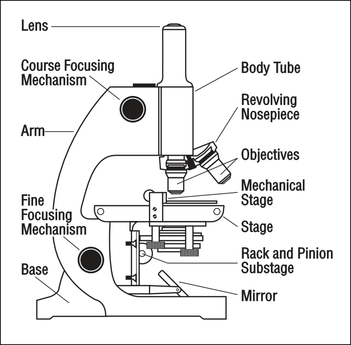
38 image of microscope with label
Microscope Labeling - The Biology Corner Students label the parts of the microscope in this photo of a basic laboratory light microscope. Can be used for practice or as a quiz. ... Microscope Labeling . Microscope Use: 15. When focusing a specimen, you should always start with the _____ objective. 16. When using the high power objective, only the _____ knob should be used. 17. The ... Compound Microscope - Diagram (Parts labelled), Principle and Uses See: Labeled Diagram showing differences between compound and simple microscope parts Structural Components The three structural components include 1. Head This is the upper part of the microscope that houses the optical parts 2. Arm This part connects the head with the base and provides stability to the microscope.
Compound Microscope Parts - Labeled Diagram and their Functions - Rs ... The eyepiece (or ocular lens) is the lens part at the top of a microscope that the viewer looks through. The standard eyepiece has a magnification of 10x. You may exchange with an optional eyepiece ranging from 5x - 30x. [In this figure] The structure inside an eyepiece. The current design of the eyepiece is no longer a single convex lens.

Image of microscope with label
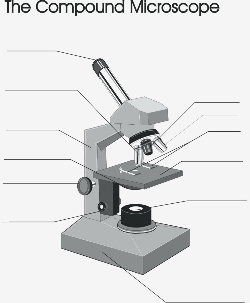
Microscope Parts and Functions Microscope Parts and Functions With Labeled Diagram and Functions How does a Compound Microscope Work?. Before exploring microscope parts and functions, you should probably understand that the compound light microscope is more complicated than just a microscope with more than one lens.. First, the purpose of a microscope is to magnify a small object or to magnify the fine details of a larger ... Parts of a microscope with functions and labeled diagram Q. List down the 18 parts of a Microscope. 1. Ocular Lens (Eye Piece) 2. Diopter Adjustment 3. Head 4. Nose Piece 5. Objective Lens 6. Arm (Carrying Handle) 7. Mechanical Stage 8. Stage Clip 9. Aperture 10. Diaphragm 11. Condenser 12. Coarse Adjustment 13. Fine Adjustment 14. Illuminator (Light Source) 15. Stage Controls 16. Base 17. Compound Microscope Parts, Functions, and Labeled Diagram Compound Microscope Definitions for Labels. Eyepiece (ocular lens) with or without Pointer: The part that is looked through at the top of the compound microscope. Eyepieces typically have a magnification between 5x & 30x. Monocular or Binocular Head: Structural support that holds & connects the eyepieces to the objective lenses.
Image of microscope with label. Microscope Types (with labeled diagrams) and Functions The working principle of a simple microscope is that when a lens is held close to the eye, a virtual, magnified and erect image of a specimen is formed at the least possible distance from which a human eye can discern objects clearly. Simple microscope labeled diagram Simple microscope functions It is used in industrial applications like: Microscope Illustrations and Stock Art. 68,272 Microscope illustration ... Microscope Drawing by bruno1998 7 / 9,315 microscope Stock Illustration by get4net 7 / 818 Microscope Stock Illustrations by Orla 11 / 603 Microscope Stock Illustration by cteconsulting 5 / 1,066 Microscope Kids Drawings by lenm 11 / 3,366 Microscope Stock Illustration by cteconsulting 4 / 511 Microscope Drawings by premiumdesign 2 / 916 3d ... Parts of the Microscope with Labeling (also Free Printouts) Microscopes are specially created to magnify the image of the subject being studied. This exercise is created to be used in homes and schools. the microscope layout, including the blank and answered versions are available as pdf downloads. Click to Download : Label the Parts of the Microscope (A4) PDF print version. Compound Microscope: Definition, Diagram, Parts, Uses, Working ... - BYJUS Compound microscope is a type of optical microscope that is used for obtaining a high-resolution image. There are more than two lenses in a compound microscope. Learn about the working principle, parts and uses of a compound microscope along with a labeled diagram here.
Microscope, Microscope Parts, Labeled Diagram, and Functions Revolving Nosepiece or Turret: Turret is the part of the microscope that holds two or multiple objective lenses and helps to rotate objective lenses and also helps to easily change power. Objective Lenses: Three are 3 or 4 objective lenses on a microscope. The objective lenses almost always consist of 4x, 10x, 40x and 100x powers. The most common eyepiece lens is 10x and when it coupled with ... Microscope Labeling Game - PurposeGames.com About this Quiz. This is an online quiz called Microscope Labeling Game. There is a printable worksheet available for download here so you can take the quiz with pen and paper. This quiz has tags. Click on the tags below to find other quizzes on the same subject. Science. Parts of a Simple Microscope - Labeled (with diagrams) A simple microscope is a very first type of microscope ever created. It consists of simple parts and performs simple functions. Although there are now many advanced microscope types, some applications may still demand the use of a simple microscope. In this article, we are going to discuss the parts and functions of a simple microscope. Microscope Drawing And Label - Painting Valley Are you looking for the best images of Microscope Drawing And Label? Here you are! We collected 33+ Microscope Drawing And Label paintings in our online museum of paintings - PaintingValley.com. ADVERTISEMENT LIMITED OFFER: Get 10 free Shutterstock images - PICK10FREE label microscope diagram compound parts light labeling functions microscopic
Labeling the Parts of the Microscope Labeling the Parts of the Microscope This activity has been designed for use in homes and schools. Each microscope layout (both blank and the version with answers) are available as PDF downloads. You can view a more in-depth review of each part of the microscope here. Download the Label the Parts of the Microscope PDF printable version here. 22 Parts Of a Microscope With Their Function And Labeled Diagram A microscope is a laboratory instrument used to examine objects that are too small to be seen by the naked eye. In other words, it enlarges images of small objects. Invented by a Dutch spectacle maker in the late 16th century, light microscopes use lenses and light to magnify images. PDF Label parts of the Microscope Label parts of the Microscope: . Created Date: 20150715115425Z 300+ Free Microscope & Laboratory Images - Pixabay Upload 399 Free images of Microscope Related Images: laboratory science bacteria research scientist lab biology chemistry medical Find your perfect microscope image. Free pictures to download and use in your next project. 399 Free images of Microscope / 4‹ ›
Microscope Labeling | Microscope, Student focus, Direct instruction
Solved Label the image of a compound light microscope using - Chegg Experts are tested by Chegg as specialists in their subject area. We review their content and use your feedback to keep the quality high. Transcribed image text: Label the image of a compound light microscope using the terms provided.
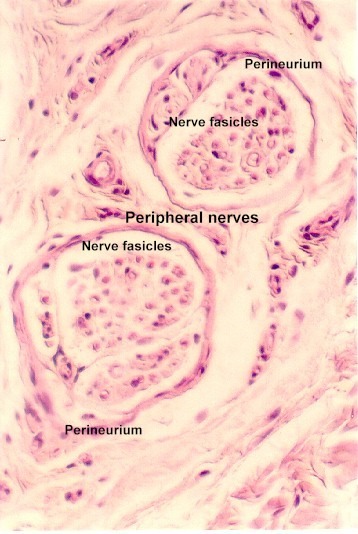
Peripheral Nerve Histology | Microanatomy Web Atlas | Gwen V. Childs, Ph.D.
Simple Microscope - Parts, Functions, Diagram and Labelling Simple Microscope - Parts, Functions, Diagram and Labelling A microscope is one of the commonly used equipment in a laboratory setting. A microscope is an optical instrument used to magnify an image of a tiny object; objects that are not visible to the human eyes. Table of Contents The common types of microscopes are: What is a Simple microscope?
microscope with labels - /science/tools/microscope/microscope_with ...
Skin Images Labeled | Virtual Anatomy Lab VAL - ncccval Skin Images Labeled | Virtual Anatomy Lab VAL ... Connective Tissue Images Unlabeled. Microscope. Microscope Images Labeled. Microscope Images Unlabeled. Mitosis. Mitosis Images Labeled. Mitosis Images Unlabeled. Skin. Skin Images Labeled. Skin Images Unlabeled. Skeletal system. Skeletal Images Labeled. Skeletal Images Unlabeled.

Microscope Labeled Simple - Micropedia
Compound Microscope with labels Stock Vector | Adobe Stock Download Compound Microscope with labels Stock Vector and explore similar vectors at Adobe Stock. Adobe Stock. Photos Illustrations Vectors Videos Audio Templates Free Premium Editorial Fonts. Plugins. 3D. Photos Illustrations Vectors Videos Audio Templates Free Premium Editorial Fonts.

Euglena viridis - BF, DIC microscope 400x - YouTube
Label the microscope — Science Learning Hub All microscopes share features in common. In this interactive, you can label the different parts of a microscope. Use this with the Microscope parts activity to help students identify and label the main parts of a microscope and then describe their functions. Drag and drop the text labels onto the microscope diagram.
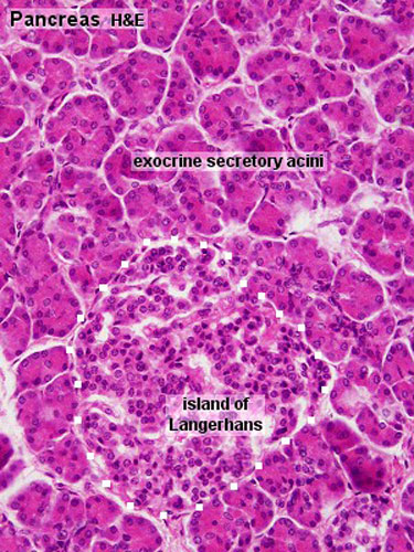
Gastrointestinal Tract - Pancreas Histology - Embryology
Microscope Parts, Function, & Labeled Diagram - slidingmotion Condenser. The condenser is to focus the light, which passes from the microscopic illuminator to the specimen. This condenser is located just below the diaphragm. This diaphragm is one of the important parts of the compound microscope which will help to get an accurate and sharp image. The condenser has a magnification power of 400X and above.

32 Label Of Compound Microscope - Label Design Ideas 2020
Microscope Labeling - The Biology Corner The labeling worksheet could be used as a quiz or as part of direct instruction where students label the microscope as you go over what each part is used for. The google slides shown below have the same microscope image with the labels for students to copy. I often spend the first day walking students through the steps and having them look at a ...
Digestive
Microscope picture label Flashcards | Quizlet Start studying Microscope picture label. Learn vocabulary, terms, and more with flashcards, games, and other study tools.

Labeling a Compound Microscope Quiz
Parts of a Compound Microscope - Labeled (with diagrams) It is used to carry the microscope and at the same time connect the base of the microscope to the head. (1, 2, 3, and 4) Image 3: A compound microscope with a corresponding label of the different parts.

Labels Of The Microscope
Microscope Labeled Pictures, Images and Stock Photos Browse 48 microscope labeled stock photos and images available, or start a new search to explore more stock photos and images. Newest results Fluorescent Imaging immunofluorescence of cancer cells growing... Plant Tissue Systems vector illustration. Labeled biology... Microscope diagram vector illustration. Labeled zoom instrument...

Rhizopus sporangia | Microbiology lab, Microbiology study, Microbiology
Compound Microscope- Definition, Labeled Diagram, Principle, Parts, Uses In order to ascertain the total magnification when viewing an image with a compound light microscope, take the power of the objective lens which is at 4x, 10x or 40x and multiply it by the power of the eyepiece which is typically 10x. Therefore, a 10x eyepiece used with a 40X objective lens will produce a magnification of 400X.

Microscope labeling
Compound Microscope Parts, Functions, and Labeled Diagram Compound Microscope Definitions for Labels. Eyepiece (ocular lens) with or without Pointer: The part that is looked through at the top of the compound microscope. Eyepieces typically have a magnification between 5x & 30x. Monocular or Binocular Head: Structural support that holds & connects the eyepieces to the objective lenses.

Electron Microscopy introduction - WUR
Parts of a microscope with functions and labeled diagram Q. List down the 18 parts of a Microscope. 1. Ocular Lens (Eye Piece) 2. Diopter Adjustment 3. Head 4. Nose Piece 5. Objective Lens 6. Arm (Carrying Handle) 7. Mechanical Stage 8. Stage Clip 9. Aperture 10. Diaphragm 11. Condenser 12. Coarse Adjustment 13. Fine Adjustment 14. Illuminator (Light Source) 15. Stage Controls 16. Base 17.
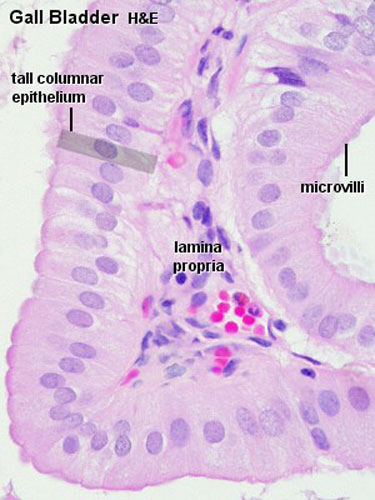
Gastrointestinal Tract - Gallbladder Histology - Embryology
Microscope Parts and Functions Microscope Parts and Functions With Labeled Diagram and Functions How does a Compound Microscope Work?. Before exploring microscope parts and functions, you should probably understand that the compound light microscope is more complicated than just a microscope with more than one lens.. First, the purpose of a microscope is to magnify a small object or to magnify the fine details of a larger ...
29 You Will Love Labeling A Compound Microscope — db-excel.com
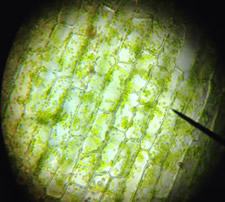
Plant Cell Lab - Onion and Elodea

Microscope labeling

Human Bio: Microscopy
Post a Comment for "38 image of microscope with label"