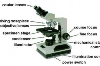
44 microscope labeling and functions
AtFTCD-L, a trans-Golgi network localized protein, modulates root ... The Golgi apparatus is an important intracellular organelle, as it performs the functions of protein modification and processing and vesicle sorting in cells (Glick et al. 2009; Papanikou and Glick 2014 ). The trans -Golgi network (TGN) is a series of interconnected tubular structures located on the trans -face of the Golgi apparatus. Microscope, Microscope Parts, Labeled Diagram, and Functions Jan 19, 2022 — Microscope Parts and Functions ; Base, Supports the microscope ; Arm, Used to carry the microscope ; Stage, Platform where the slide with the ...Microscope Parts: Microscope Parts FunctionsObjective lenses: Low-, medium-, and high-po...Light source: Provides light for viewing the spe...Base: Supports the microscope
Hanging Drop Method for Bacterial Motility - Microbe Online Hanging Drop Method Preparation. Take a clean glass slide and apply a paraffin ring, adhesive-tape ring to make circular concavity. (This step is not needed if a glass slide with depression is available). Hold a clean coverslip by its edges and carefully dab vaseline on its corners using a toothpick. Place a loopful of the fresh broth culture ...

Microscope labeling and functions
en.wikipedia.org › wiki › Electron_microscopeElectron microscope - Wikipedia An electron microscope is a microscope that uses a beam of accelerated electrons as a source of illumination. As the wavelength of an electron can be up to 100,000 times shorter than that of visible light photons , electron microscopes have a higher resolving power than light microscopes and can reveal the structure of smaller objects. Light Microscope (Theory) - Amrita Vishwa Vidyapeetham Optical microscopes function on the basis of optical theory of lenses by which it can magnifies the image obtained by the movement of a wave through the sample. The waves used in optical microscopes are electromagnetic and that in electron microscopes are electron beams. Next-Generation Laser Scanning Multiphoton Microscopes are Turnkey ... The Prospective Instruments MPX-series is a turnkey compact multimodal microscope enabling advanced multiphoton imaging without requiring a laboratory or optical bench. One of the key design features is the ultrafast fiber laser engine integrated into the free moving scan head, which is lightweight ...
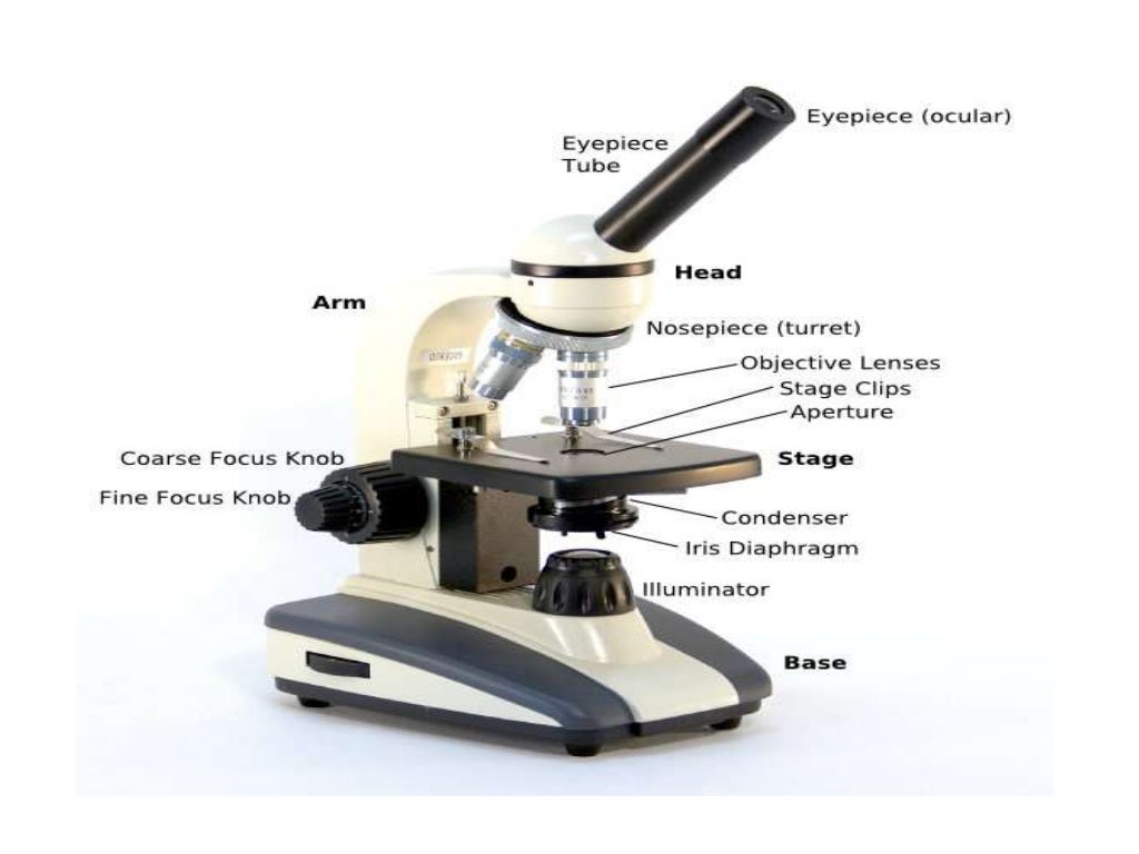
Microscope labeling and functions. Parts of a microscope with functions and labeled diagram Apr 19, 2022 — Optical parts of a microscope and their functions · Eyepiece – also known as the ocular. · Eyepiece tube – it's the eyepiece holder. · Objective ...Diagrammatically, identify the various parts of a microscope.List down the 18 parts of a Microscope. thebiologynotes.com › microscopeMicroscope- Definition, Parts, Functions, Types, Diagram, Uses Feb 21, 2022 · Limitations of Dissecting Microscope or Stereo Microscope. Has limited use; Low magnification; Costly system; 8. Digital Microscope. Digital Microscope is a type of microscope that lack an ocular lens and instead contains a digital camera and screen to display image digitally. This is a modern microscope which is a computerized system combining ... › products › microscopeMicroscope Imaging Software | Products | Leica Microsystems Jun 15, 2021 · Microscope Imaging Software. Microscope imaging software from Leica Microsystems combines microscope, digital camera and accessories into one fully integrated solution. With an intuitive user interface and straightforward navigation, it guides the user through any workflow, whether fast image acquisition or sophisticated expert analysis. ANSWER ALL THE MULTIPLE CHOICE QUESTIONS Organization of the cell, the ... ANSWER ALL THE MULTIPLE CHOICE QUESTIONS Organization of the cell, the basic unit of life, its organization critical to its ability to carry out all life activities and will learn whom the shape and size are adapted for its function. The cell is under the
› products › microscopeLAS X Life Science Microscope Software | Products | Leica ... Microscope Software Platform LAS X Life Science Do you struggle with a variety of instruments, protocols, and software interfaces in your lab? Leica Application Suite X (LAS X) is the one software platform for all Leica microscopes: It integrates confocal, widefield, stereo, super-resolution, and light-sheet instruments from Leica Microsystems. › articles › s41596/020/0399-0Proximity labeling in mammalian cells with TurboID and split ... Nov 02, 2020 · In HEK293T cells, TurboID labeling can be detected with 1–10 min of labeling, and split-TurboID labeling can be detected with 0.5–4 h of labeling 6,7. Negative controls that omit the ligase or ... abberior-instruments.com › products › minfluxMINFLUX | Abberior Instruments With its revolutionary MINFLUX microscope, abberior is raising the bar for molecular tracking with world-record temporal resolution, opening new doors for life-scientists across all disciplines. Choose between MINFLUX molecular imaging and tracking at the push of a button! Transport of intensity diffraction tomography with non-interferometric ... Since its invention in the 1600s, the optical microscope has experienced continuous development and become an indispensable tool for the visualization of micro-scale objects with high resolution in...
EOF › game › microscope-labelingMicroscope Labeling Game - PurposeGames.com This is an online quiz called Microscope Labeling Game. There is a printable worksheet available for download here so you can take the quiz with pen and paper. This quiz has tags. Click on the tags below to find other quizzes on the same subject. Gram Stain Technique - Amrita Vishwa Vidyapeetham Drawing a circle on the underside of the slide using a glassware-marking pen may be helpful to clearly designate the area in which you will prepare the smear. You may also label the slide with the initials of the name of the organism on the edge of the slide. Care should be taken that the label should not be in contact with the staining reagents. 5 White Blood Cells Types and Their Functions - New Health Advisor Function: Eosinophils work by releasing toxins from their granules to kill pathogens. The main pathogens eosinophils act against are parasites and worms. High eosinophil counts are associated with allergic reactions. 5. Basophils Basophils are the least frequent type of white blood cell, with only 0-100 cells per mm 3 of blood.
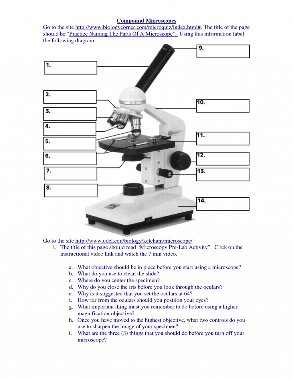
Labeled Microscope Worksheet Answers - Thekidsworksheet
KEYENCE TV : VK-X Series | KEYENCE America 3D Laser Scanning Confocal Microscope. The VK-X Series 3D Laser Scanning Confocal Microscope expands the capabilities of laser microscopy. Combining features of an optical microscope, roughness gauge, laser profilometer, and scanning electron microscope, our laser scanning microscope performs non-contact surface profile, surface roughness, and thickness measurements without the need for sample ...

Molecular make up of cells | Cells: the basic units of life | Siyavula
Next-Generation Laser Scanning Multiphoton Microscopes are Turnkey ... The Prospective Instruments MPX-series is a turnkey compact multimodal microscope enabling advanced multiphoton imaging without requiring a laboratory or optical bench. One of the key design features is the ultrafast fiber laser engine integrated into the free moving scan head, which is lightweight ...
microscope labeled : Biological Science Picture Directory – Pulpbits.net
Light Microscope (Theory) - Amrita Vishwa Vidyapeetham Optical microscopes function on the basis of optical theory of lenses by which it can magnifies the image obtained by the movement of a wave through the sample. The waves used in optical microscopes are electromagnetic and that in electron microscopes are electron beams.
Microscope parts and functions
en.wikipedia.org › wiki › Electron_microscopeElectron microscope - Wikipedia An electron microscope is a microscope that uses a beam of accelerated electrons as a source of illumination. As the wavelength of an electron can be up to 100,000 times shorter than that of visible light photons , electron microscopes have a higher resolving power than light microscopes and can reveal the structure of smaller objects.

Compound Light Microscope Labeled - Made By Creative Label
Compound Microscopes : Biological Science Picture Directory – Pulpbits.net
Microscope Review.wmv - YouTube

Mr. Klein's Classes - science_handbook_4.3 | Science teaching resources ...

# 74 Blood cells - structure and functions | Biology Notes for IGCSE 2014
Post a Comment for "44 microscope labeling and functions"