40 label this transmission electron micrograph
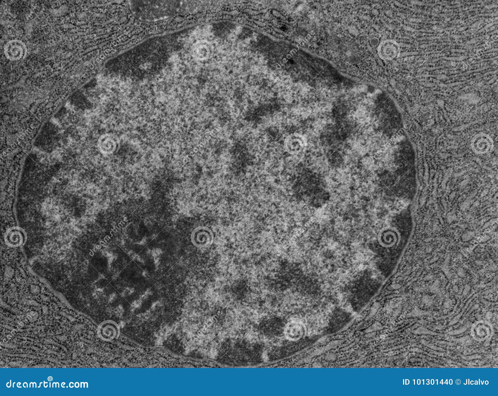
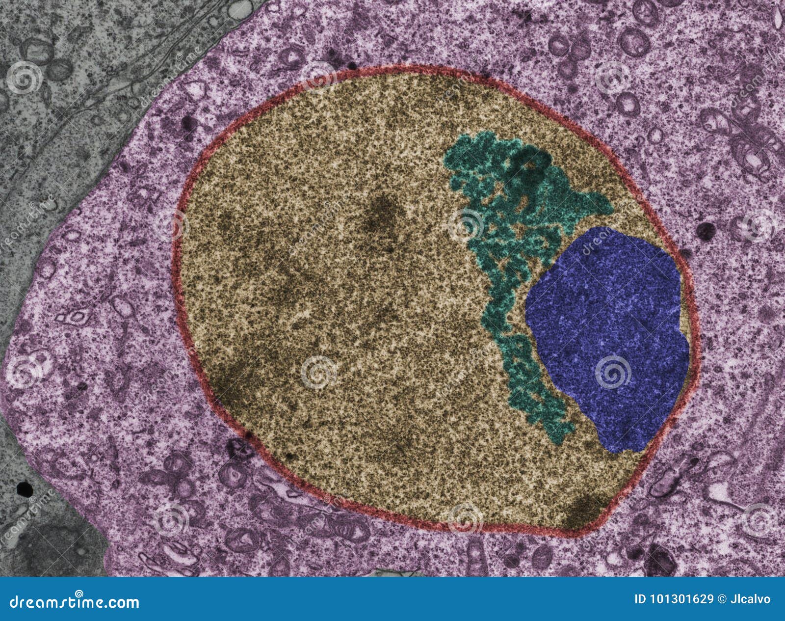
anatomy 10.png - Label the transmission electron micrograph of the ... anatomy 10.png - Label the transmission electron micrograph of the. anatomy 10.png - Label the transmission electron micrograph... School Utah Valley University; Course Title ZOOL 1090; Uploaded By emileeroylance19. Pages 1 Ratings 67% (3) 2 out of 3 people found this document helpful; Solved Label the transmission electron micrograph based on | Chegg.com Expert Answer nucleus is the house of the genetic material which contains all the h … View the full answer Transcribed image text: Label the transmission electron micrograph based on the hints provided Mitochondrion Heterochromatin Plasma cell Nucleus Rough endoplasmic reticulum Nucleolus Previous question Next question
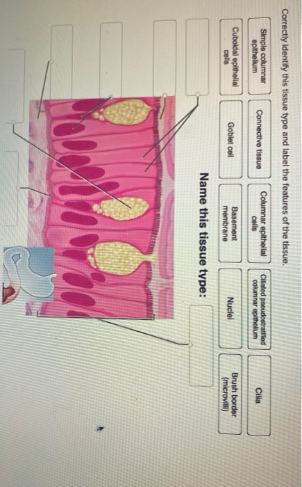
Solved Label this transmission electron micrograph of | Chegg.com Solved Label this transmission electron micrograph of | Chegg.com Science Anatomy and Physiology Anatomy and Physiology questions and answers Label this transmission electron micrograph of relaxed sarcomeres by clicking and dragging the labels to the correct location Sarcamere 1 band (light) Z disc Mline Aband (dark) H zone
Label this transmission electron micrograph
Solved Label the transmission electron micrograph of the | Chegg.com Transcribed image text: Label the transmission electron micrograph of the cell. 0 Nucleus rences Mitochondrion Heterochromatin Peroxisome Vesicle ULAR bumit Click and drag each label into the correct category to indicate whether it pertains to the cytoplasm or the plasma membrane. Nanoscale imaging of phonon dynamics by electron microscopy Jun 08, 2022 · Although dipole scattering in polar materials, such as BN 21,26,27, MgO 23 and SiC 20,28, under on-axis scanning transmission electron microscopy-electron energy loss spectroscopy (STEM-EELS) the ... Label This Transmission Electron Micrograph Of A Relaxed Sarcomere ... (b) section through a muscle in the extended condition (140 % of whole muscle resting length) label this transmission electron micrograph. Provide the labels for the electron micrograph in figure 12.8. Provide the labels for the electron micrograph in figure 18.5. Note how the sarcomeres are extended to only approximately 120 % .
Label this transmission electron micrograph. (Get Answer) - Label this transmission electron micrograph of relaxed ... Label this transmission electron micrograph of relaxed sarcomeres by clicking and dragging the labels to the correct location Sarcamere 1 band (light) Z disc Mline Aband (dark) H zone Mar 29 2022 11:37 AM Expert's Answer Solution.pdf Next Previous Q: Q: Q: Q: Posted one year ago Recent Questions in Physics Q: 1. Hepatitis A - Wikipedia Hepatitis A is an infectious disease of the liver caused by Hepatovirus A (HAV); it is a type of viral hepatitis. Many cases have few or no symptoms, especially in the young. The time between infection and symptoms, in those who develop them, is 2–6 weeks. When symptoms occur, they typically last 8 weeks and may include nausea, vomiting, diarrhea, jaundice, fever, and … Transmission electron microscopy DNA sequencing - Wikipedia Transmission electron microscopy DNA sequencing is a single-molecule sequencing technology that uses transmission electron microscopy techniques. The method was conceived and developed in the 1960s and 70s, but lost favor when the extent of damage to the sample was recognized. In order for DNA to be clearly visualized under an electron microscope, it must be labeled with heavy atoms. Immune response: MedlinePlus Medical Encyclopedia INFLAMMATION. The inflammatory response (inflammation) occurs when tissues are injured by bacteria, trauma, toxins, heat, or any other cause. The damaged cells release chemicals including histamine, bradykinin, and prostaglandins.
PDF Lecture 14 - soest.hawaii.edu Transmission electron microscopy (TEM) is a microscopy technique whereby a beam of electrons is transmitted through an ultra thin specimen, interacting with the specimen as it passes through. Microbiology (EXAM #1) CH. 3 Homework & Reading Questions A. Beams of electrons are already in the chamber before the specimen is placed; they then will react to the movement. B. They randomly shoot beams of electrons at the specimen. C. Beams of electrons that are focused by means of a magnetic field act as waves. D. These microscopes do not use beams of electrons. Instead, they use photons. Transmission electron microscopy characterization of ... - PubMed Transmission electron microscopy characterization of fluorescently labelled amyloid β 1-40 and α-synuclein aggregates BMC Biotechnol. 2011 Dec 19; 11:125. doi ... However, the use of these labels may interfere with the formation of larger-scale protein structures such as amyloid aggregates. Therefore, we investigate the effects of some ... PDF Identifying Organelles from an Electron Micrograph The photograph shown below details chloroplast structure as viewed with a transmission electron microscope Courtesy of Dr. Julian Thorpe - EM & FACS Lab, Biological Sciences University Of Sussex A single Granum Chloroplast envelope visible as two membranes Stroma containing numerous small ribosomes Lamellae connecting different grana
human eye, light microscopy , scanning electron microscopy, transmission electron microscopy , x-ray crystallography. Match each image with the type of microscopy used to produce it. ... unequal electron sharing Correct label: polar covalent bond partial charge interactions Correct label: hydrogen charge attractions Correct label: Ternary Phase Diagram - an overview | ScienceDirect Topics Ternary phase diagrams are used to represent all possible mixtures of three solvents [1]; they are described in Chapter 3.Here, we shall indicate how they should be used to minimize the solvent consumption. Figure 2.1 (top) shows the methanol–chloroform–water ternary phase diagram with the tie-lines in the biphasic domain. Five particular compositions are shown in the … Fluorescence microscope - Wikipedia This allows one to visualize ultrastructure and contextual information with the electron microscope while using the data from the fluorescence microscope as a labelling tool. [11] The first technique to really achieve a sub-diffraction resolution was STED microscopy , proposed in 1994. 6.1 Viruses - Microbiology | OpenStax Figure 6.3 (a) In this transmission electron micrograph, a bacteriophage (a virus that infects bacteria) is dwarfed by the bacterial cell it infects. (b) An illustration of the bacteriophage in the micrograph. (credit a: modification of work by U.S. Department of Energy, Office of Science, LBL, PBD)
108 Tem Showing Photos - Free & Royalty-Free Stock Photos ...
Label This Transmission Electron Micrograph / Microscopy Innovations ... Label the transmission electron micrograph of the cell. Label the transmission electron micrograph of the. File Figure 04 04 02 New Jpg Wikimedia Commons from upload.wikimedia.org Nuclear envelope nucleolus nucleus heterochromatin reset zoom · this problem has been solved . Label the transmission electron micrograph of the nucleus.

anatomy 10.png - Label the transmission electron micrograph ...
Transmission Electron Microscopy (TEM) - Warwick TEM. The transmission electron microscope is a very powerful tool for material science. A high energy beam of electrons is shone through a very thin sample, and the interactions between the electrons and the atoms can be used to observe features such as the crystal structure and features in the structure like dislocations and grain boundaries.

Overview of the data and gold standard labels for SEM (a) and ...
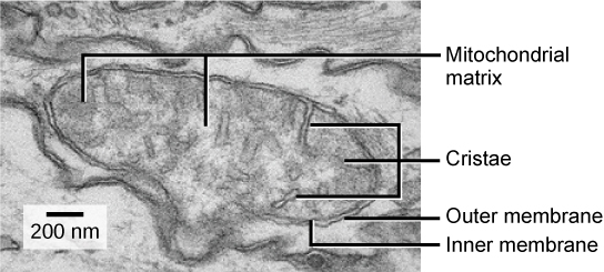
Solved Label the transmission electron micrograph of the | Chegg.com Explanation - Mitochondrion is filamentous or globular in shape, occur in variable numbers from a few hundred to few thousands in different cells. It … View the full answer Transcribed image text: Label the transmission electron micrograph of the mitochondrion. Matrix granule Mitochondrion Outer membrane Cristae Inner membrane Matrix Reset Zoom
Solved Label the transmission electron micrograph of the ...
Label This Transmission Electron Micrograph : TEM of chloroplast from ... Label this transmission electron micrograph of relaxed sarcomeres by clicking and dragging the labels to the correct location . Transmission electron microscopy (tem) is one of the oldest technologies and still. Molecular labeling for correlative microscopy: Fluorescence microscopy in combination with tem and an ion beam analysis (iba, which ...

Labeling the Cell Flashcards | Quizlet
Transmission Electron Microscope (With Diagram) Finally, the electrons are focused by an electromagnetic projector lens (instead of an ocular lens as in a light microscope) on a screen or photographic plate. The final image in a TEM is known as transmission electron micrograph. The salts of some heavy metals, e.g., lead; osmium, tungsten and uranium are often used for staining.
Solved You have not seen this stain (or color enhancement ...
Cryo-electron microscopy: A primer for the non-microscopist Over the past decade, the phrase “cryo-electron microscopy”, often abbreviated as “cryo-EM”, has evolved to encompass a broad range of experimental methods. At the core, each of these is based upon the principle of imaging radiation-sensitive specimens in a transmission electron microscope under cryogenic conditions.
Sex Vesicle. False Colour TEM Stock Image - Image of testicle ...
The Transmission Electron Microscope | CCBER Transmission electron microscopes (TEM) are microscopes that use a particle beam of electrons to visualize specimens and generate a highly-magnified image. TEMs can magnify objects up to 2 million times. In order to get a better idea of just how small that is, think of how small a cell is.

a) Bright-field cross-sectional transmission electron ...
Symmetry-breaking-induced plasmonic exceptional points … Feb 17, 2020 · The structures are fabricated using two-step electron ... presents a top-view scanning electron micrograph of the ... H. X. et al. Exceptional points in extraordinary optical transmission through ...
A tour of the cell: View as single page
Label the transmission electron micrograph of the nucleus. Label this transmission electron micrograph of relaxed sarcomeres by clicking and dragging the labels to the correct location Sarcamere 1 band (light) Z disc Mline Aband (dark) H zone Posted 2 months ago. Recent Questions in Economics - Others . Q: Question 1a Assume you are the Director of Marketing for Majjus Enterprise, a firm that produces ...

Transmission electron micrographs of the ink-release vesicle ...
The Effect of Electron Beam Irradiation in Environmental Scanning ... Figure 1 Illustration of environmental scanning electron microscopy of whole eukaryotic cells in liquid using scanning transmission electron microscopic detection. Cells grown on a silicon microchip with thin silicon nitride (SiN) windows are maintained in liquid and contrast is obtained on nanoparticles attached to the desired proteins [epidermal growth factor receptors (EGFR)].
9700 QR Dynamic Papers Biology al Cambridge
Transmission Electron Microscopy: study guides and answers on ... - Quizlet E) a phase-contrast light microscope. a transmission electronic microscope. The advantage of light microscopy over electron microscopy is that A) light microscopy allows one to view dynamic processes in living cells. B) light microscopy provides for higher resolving power than electron microscopy.

Immunogold-labeling of glutathione. Transmission electron ...
Electron Micrographs** Electron Micrographs**. Below is a collection of electron micrographs with labelled subcellular structures that you should be able to identify. Also, be sure to observe any electron micrographs which are made available in the laboratory by the instructor. You should concentrate on the similarities in form that permit identification of the ...

Solved Label the transmission electron micrograph of the ...
HIV/AIDS - Wikipedia Acquired immunodeficiency syndrome (AIDS) is defined as an HIV infection with either a CD4 + T cell count below 200 cells per µL or the occurrence of specific diseases associated with HIV infection. In the absence of specific treatment, around half of people infected with HIV develop AIDS within ten years. The most common initial conditions that alert to the presence of AIDS …

Transmission electron micrographs of chloroplasts and ...
Imaging Specific Protein Labels on Eukaryotic Cells in Liquid with ... Figure 1: The principle of liquid scanning transmission electron microscopy (STEM). Cells kept in liquid are enclosed between two electron-transparent silicon nitride windows. Scanning a focused electron beam over the sample leads to the detection of elastically scattered electrons with an annular dark field detector.

Figure, Transmission Electron Micrograph of Rough Endoplasmic ...
IMG_2132.jpg - FIGURES Label this transmission electron micrograph ( 16 ... SCIENCE 101 IMG_2132.jpg - FIGURES Label this transmission electron micrograph ( 16, 000 X ) of a relaxed sarcomere by placing the correct numbers in the spaces IMG_2132.jpg - FIGURES Label this transmission electron... School University High School, Tucson Course Title SCIENCE 101 Type Homework Help Uploaded By mmederos254 Pages 1 Ratings 100% (1)
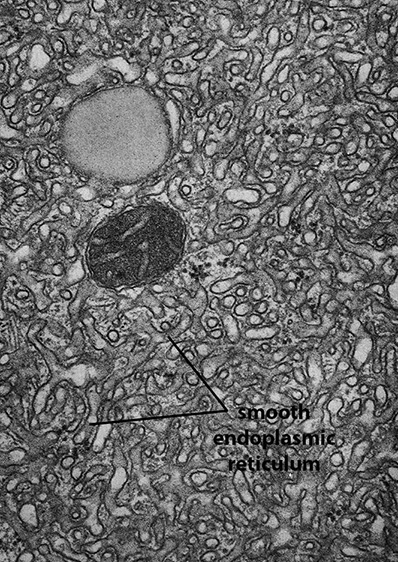
BIOL 230 Lecture Guide - Electron Micrograph of Rough ...
Solved Label the transmission electron micrograph of the | Chegg.com Expert Answer Answer The label is indicated from TOP to BOTTOM Ciliu … View the full answer Transcribed image text: Label the transmission electron micrograph of the cilium. Microvillus Axoneme Cilium Dynein arm Previous question Next question
Nanomaterials | Free Full-Text | A Guide for Using ...
Uniform nucleation and epitaxy of bilayer molybdenum disulfide 04/05/2022 · Figure 5i shows an optical micrograph of adjacent bilayer MoS 2 domains with the same orientation. However, their SHG intensity was …
2.3.3 Identify structures from electron micrographs of liver ...
Q&A: What are exosomes, exactly? - PMC 13/06/2016 · Exosomes correspond to intraluminal vesicles of multivesicular bodies. A transmission electron micrograph of an Epstein–Barr virus-transformed B cell displaying newly expelled exosomes at the plasma membrane. ... Electron microscopy of isolated fractions as ‘whole mounts’ make it possible to immuno-label vesicles, with the limitation that ...

anatomy 10.png - Label the transmission electron micrograph ...
Transmission electron microscopy DNA sequencing - Google Transmission electron microscopy DNA sequencing is a single-molecule sequencing technology that uses transmission electron microscopy techniques.The method was conceived and developed in the 1960s and 70s, but lost favor when the extent of damage to the sample was recognized. DNA is visible under the electron microscope; however, it must be labeled with heavy atoms so that the DNA bases can be ...

Immunogold labelling - Wikipedia
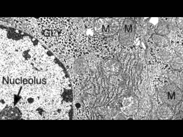
Electron Micrographs of Cell Organelles | Zoology This is an electron micrograph of nucleus. (Fig. 17 & 18): (1) Nucleus was discovered by Brown (1831). (2) It is a characteristic entity of almost all eukaryotic cells except mammalian RBCs. (3) The nucleus is generally one but may also be two, four or many.

Transmission electron micrograph of a gold-labelled Lowicryl ...
Labeling the Cell Flashcards | Quizlet Label the transmission electron micrograph of the nucleus. membrane bound organelles golgi apparatus, mitochondrion, lysosome, peroxisome, rough endoplasmic reticulum nonmembrane bound organelles ribosomes, centrosome, proteasomes cytoskeleton includes microfilaments, intermediate filaments, microtubules Identify the highlighted structures

Differentiation of Naturally Produced Extracellular ...
Label This Transmission Electron Micrograph Of A Relaxed Sarcomere ... (b) section through a muscle in the extended condition (140 % of whole muscle resting length) label this transmission electron micrograph. Provide the labels for the electron micrograph in figure 12.8. Provide the labels for the electron micrograph in figure 18.5. Note how the sarcomeres are extended to only approximately 120 % .

a) A high-resolution transmission electron micrograph of ...
Nanoscale imaging of phonon dynamics by electron microscopy Jun 08, 2022 · Although dipole scattering in polar materials, such as BN 21,26,27, MgO 23 and SiC 20,28, under on-axis scanning transmission electron microscopy-electron energy loss spectroscopy (STEM-EELS) the ...

Transmission electron microscopy (TEM) micrograph of ...
Solved Label the transmission electron micrograph of the | Chegg.com Transcribed image text: Label the transmission electron micrograph of the cell. 0 Nucleus rences Mitochondrion Heterochromatin Peroxisome Vesicle ULAR bumit Click and drag each label into the correct category to indicate whether it pertains to the cytoplasm or the plasma membrane.
Reading: Mitochondria | Biology (Early Release) | | Course Hero
Solved Label the transmission electron micrograph of the ...

Chapter 14 & 15 Flashcards Flashcards | Quizlet

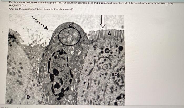
Transmission electron micrograph (TEM) of the epithelium in ...

Transmission electron micrograph of an Encephalitozoon ...

Transmission electron micrographs of mesophyll cells among ...

Transmission electron micrograph of turkey spermatozoa ...
Solved A TEME is a transmission electron micro taken with a ...
1 IB Biology Higher Level Summer Assignment All parts of your ...

Dynamics of mitochondrial cristae. (a) Transmission electron ...
CcFIG 5 Legend

lab 12 word.docx - Name: Laboratory Assessment Date: 12 ...

Transmission electron microscopy images of immunogold-labeled ...

Transmission electron micrograph of an animal cell - Stock ...

Get Answer) - Label this transmission electron micrograph of ...
Microscopy Innovations | Transmission electron microscopy (TEM)
Post a Comment for "40 label this transmission electron micrograph"