38 draw and label the microscope
Labeling the Parts of the Microscope | Microscope activity, Science ... Jan 13, 2016 - Free worksheets for labeling parts of the microscope including a worksheet that is blank and one with answers. A Study of the Microscope and its Functions With a Labeled Diagram A Study of the Microscope and its Functions With a Labeled Diagram To better understand the structure and function of a microscope, we need to take a look at the labeled microscope diagrams of the compound and electron microscope. These diagrams clearly explain the functioning of the microscopes along with their respective parts.
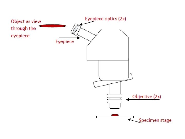
Simple Microscope - Diagram (Parts labelled), Principle, Formula and Uses The working principle of a simple microscope is that when a lens is held close to the eye, a virtual, magnified and erect image of a specimen is formed at the least possible distance from which a human eye can discern objects clearly. Magnification formula The magnification power of a simple microscope is expressed as: M = 1 + D/F Where
Draw and label the microscope
How To Draw A Microscope - YouTube Today, we're learning how to draw a cool microscope!👩🎨 JOIN OUR ART HUB MEMBERSHIP! VISIT 🎨 VISIT OUR AMAZON ART SUPPLY S... Microscope Parts and Functions First, the purpose of a microscope is to magnify a small object or to magnify the fine details of a larger object in order to examine minute specimens that cannot be seen by the naked eye. Here are the important compound microscope parts... Eyepiece: The lens the viewer looks through to see the specimen. Light Microscope- Definition, Principle, Types, Parts, Labeled Diagram ... A light microscope is a biology laboratory instrument or tool, that uses visible light to detect and magnify very small objects and enlarge them. They use lenses to focus light on the specimen, magnifying it thus producing an image. The specimen is normally placed close to the microscopic lens.
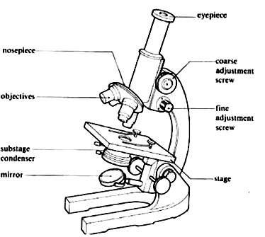
Draw and label the microscope. Label the microscope — Science Learning Hub Drag and drop the text labels onto the microscope diagram. If you want to redo an answer, click on the box and the answer will go back to the top so you can move it to another box. If you want to check your answers, use the Reset incorrect button. This will reset incorrect answers only. Microscope Labeling Diagram | Quizlet Unit 2 Lesson 5 - Punnett Squares and Pedigrees. 4 terms. PGFry210. Unit 2 Lesson 4 - Heredity. 9 terms. PGFry210. Upgrade to remove ads. Only $2.99/month. Compound Microscope- Definition, Labeled Diagram, Principle, Parts, Uses The naked eye can now view the specimen at magnification 400 times greater and so microscopic details are revealed. Alternatively, the magnification of the compound microscope is given by: m = D/ fo * L/fe where, D = Least distance of distinct vision (25 cm) L = Length of the microscope tube fo = Focal length of the objective lens Compound Microscope Parts - Labeled Diagram and their Functions - Rs ... Labeled diagram of a compound microscope Major structural parts of a compound microscope There are three major structural parts of a compound microscope. The head includes the upper part of the microscope, which houses the most critical optical components, and the eyepiece tube of the microscope.
Parts of a Microscope Labeling Activity - Storyboard That Create a poster that labels the parts of a microscope and includes descriptions of what each part does. Click "Start Assignment". Use a landscape poster layout (large or small). Search for a diagram of a microscope. Using arrows and textables label each part of the microscope and describe its function. How to Sketch a Microscope Slide - Identifying and Sketching Cell ... First, to represent the microscope field of view, draw a circle on the page - this can be freehand or, if you want to be precise, use a compass. If you are using a graticule slide (a microscope slide with millimeter grid lines), lightly sketch a grid over your circle. Label The Parts Of A Microscope Teaching Resources | TpT Hashtag Teached. 4.9. (10) $3.00. $2.00. PDF. Check out this well-organized Microscope label and describe worksheet. Part I is a visual that students will label and it corresponds with Part II where they will describe the function of those parts. This is a great worksheet that can easily be used for classwork, group work, homework, assessments ... Microscope Labeling - The Biology Corner Students label the parts of the microscope in this photo of a basic laboratory light microscope. Can be used for practice or as a quiz. ... Microscope Labeling . Microscope Use: 15. When focusing a specimen, you should always start with the _____ objective. 16. When using the high power objective, only the _____ knob should be used. 17. The ...
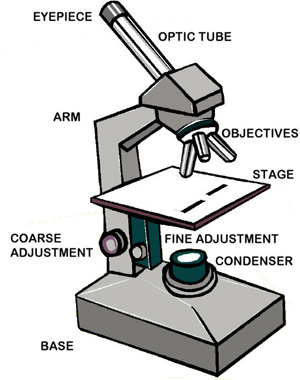
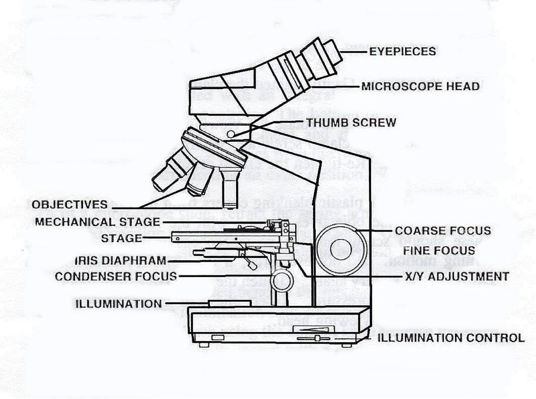
Label Microscope Diagram - EnchantedLearning.com Using the terms listed below, label the microscope diagram. arm - this attaches the eyepiece and body tube to the base. base - this supports the microscope. body tube - the tube that supports the eyepiece. coarse focus adjustment - a knob that makes large adjustments to the focus. diaphragm - an adjustable opening under the stage, allowing ... Microscope Drawing Easy with Label - YouTube In this video I go over a microscope drawing that is easy with label. There is a blank copy at the end of the video to review on your own. A great way to s... Simple Microscope - Parts, Functions, Diagram and Labelling Stage - The stage of the microscope is a metal plate that is rectangular in shape and fitted to the vertical rod. It comes with a hole in the center that enables the light to pass from below. The stage holds the slide that contains the specimen to be examined for. Microscope Drawing: How to Sketch Microscope Slides How to Draw Microscope Slides Organize and orient your field of view: To begin, draw a circle as large as possible with a pencil. An 8.5 x 11-inch piece of paper is good size for beginners. The circle represents what you see through the eyepiece of the microscope. Using thin lines, divide the circle into quarters in order to organize the picture.
How to Draw a Microscope Easy
PDF Label parts of the Microscope: Answers Label parts of the Microscope: Answers Coarse Focus Fine Focus Eyepiece Arm Rack Stop Stage Clip . Created Date: 20150715115425Z ...

Label the numbered parts of the microscope - ppt download
Labeling the Parts of the Microscope Labeling the Parts of the Microscope This activity has been designed for use in homes and schools. Each microscope layout (both blank and the version with answers) are available as PDF downloads. You can view a more in-depth review of each part of the microscope here. Download the Label the Parts of the Microscope PDF printable version here.

How to draw compound of Microscope easily - step by step
Microscope Diagram and Functions | Science fair projects, Microscope ... A Study of the Microscope and its Functions With a Labeled Diagram To better understand the structure and function of a microscope, we need to take a look at the labeled microscope diagrams of the compound and electron microscope. These diagrams clearly explain the functioning of the microscopes along with their respective parts. M mooketsi

Parts of a Compound Microscope and Their Functions
Draw And Label A Microscope - Free PDF File Sharing Draw And Label A Microscope. June 6th, 2013 14:58:10 PM ... Draw and label two or three Elodea cells, including cell wall, ... Observe the protists under the microscope. 4. Draw several protists and estimate their length. [Filename: cells_microscopy.pdf] - Read File Online - Report Abuse. microscope activity - The Science Spot ...
Comparing and Contrasting the Different Parts of the Microscope
Free Microscope Worksheets for Simple Science Fun for Your Students After cutting out the words, your kids can arrange in the correct boxes to label the parts. Once your kids are certain about their decisions, they can glue onto the area. ... The Microscope includes a colorful drawing of a microscope and lines to add observations and such. Microscope Lab Report includes colorful drawings of a microscope, test ...

Label Of Microscope - ClipArt Best
Microscope Drawing Teaching Resources | Teachers Pay Teachers This GEEKS lab sheet gives students room to draw three specimens. It includes labels for the Specimen Name, Magnification, Observations/ Challenges. The last label gives students an opportunity to write down something interest them may observe, or record challenges they had while trying to observe their specimens.

Cartoon Sticker Stick Icon Decal Label Microscope Science ...
Parts of the Microscope with Labeling (also Free Printouts) Parts of the Microscope with Labeling (also Free Printouts) A microscope is one of the invaluable tools in the laboratory setting. It is used to observe things that cannot be seen by the naked eye. Table of Contents 1. Eyepiece 2. Body tube/Head 3. Turret/Nose piece 4. Objective lenses 5. Knobs (fine and coarse) 6. Stage and stage clips 7. Aperture
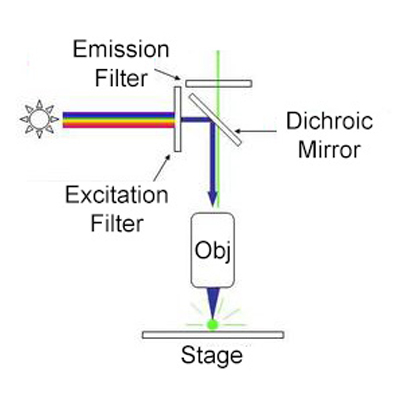
Fluorescence Microscopy - Explanation and Labelled Images ...
Microscope Parts, Function, & Labeled Diagram - slidingmotion Microscope parts labeled diagram gives us all the information about its parts and their position in the microscope. Microscope Parts Labeled Diagram The principle of the Microscope gives you an exact reason to use it. It works on the 3 principles. Magnification Resolving Power Numerical Aperture. Parts of Microscope Head Base Arm Eyepiece Lens
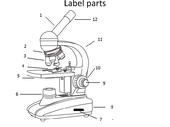
Compound Microscope Review Label parts 1 body tube
Microscope, Microscope Parts, Labeled Diagram, and Functions Microscopes magnify or enlarge small objects such as cells, microbes, bacteria, viruses, microorganisms etc. at a viewable scale for examination and analysis. Microscopes consist of one or more magnification lenses to enlarge the image of the microscopic objects placed in the focal plane.

in a long bond paper draw and label the parts of a compound ...
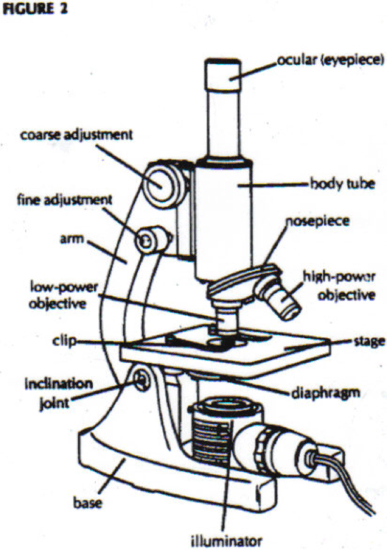
Labelled Diagram of Compound Microscope - Biology Discussion The below mentioned article provides a labelled diagram of compound microscope. Part # 1. The Stand: The stand is made up of a heavy foot which carries a curved inclinable limb or arm bearing the body tube. The foot is generally horse shoe-shaped structure (Fig. 2) which rests on table top or any other surface on which the microscope in kept.

Simple Microscope - Diagram (Parts labelled), Principle ...
How To Draw A Microscope Step by Step - [12 Easy Phase] How To Draw a Microscope Step by Step for Beginners. Pigeon. Snowman. Jeep Grand Cherokee. Horseshoe crab. Table Lamp. Rolling pin with Board. Eiffel Tower. Sauropod.

Simple Microscope Definition, Magnification, Parts And Uses
Light Microscope- Definition, Principle, Types, Parts, Labeled Diagram ... A light microscope is a biology laboratory instrument or tool, that uses visible light to detect and magnify very small objects and enlarge them. They use lenses to focus light on the specimen, magnifying it thus producing an image. The specimen is normally placed close to the microscopic lens.
LAB 1: Scientific Method/Tools of Scientific Inquiry
Microscope Parts and Functions First, the purpose of a microscope is to magnify a small object or to magnify the fine details of a larger object in order to examine minute specimens that cannot be seen by the naked eye. Here are the important compound microscope parts... Eyepiece: The lens the viewer looks through to see the specimen.

Microscope
How To Draw A Microscope - YouTube Today, we're learning how to draw a cool microscope!👩🎨 JOIN OUR ART HUB MEMBERSHIP! VISIT 🎨 VISIT OUR AMAZON ART SUPPLY S...

Parts of a Microscope - TessShaheenmicroscopy
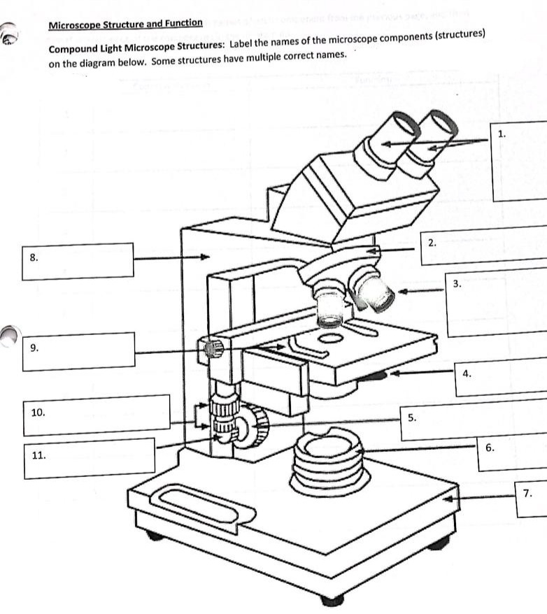
Answered: Microscope Structure and Function… | bartleby

Dissecting Stereo Microscope Parts and Functions
Microscope. Vector drawing stock vector. Illustration of ...
Parts of a microscope with functions and labeled diagram
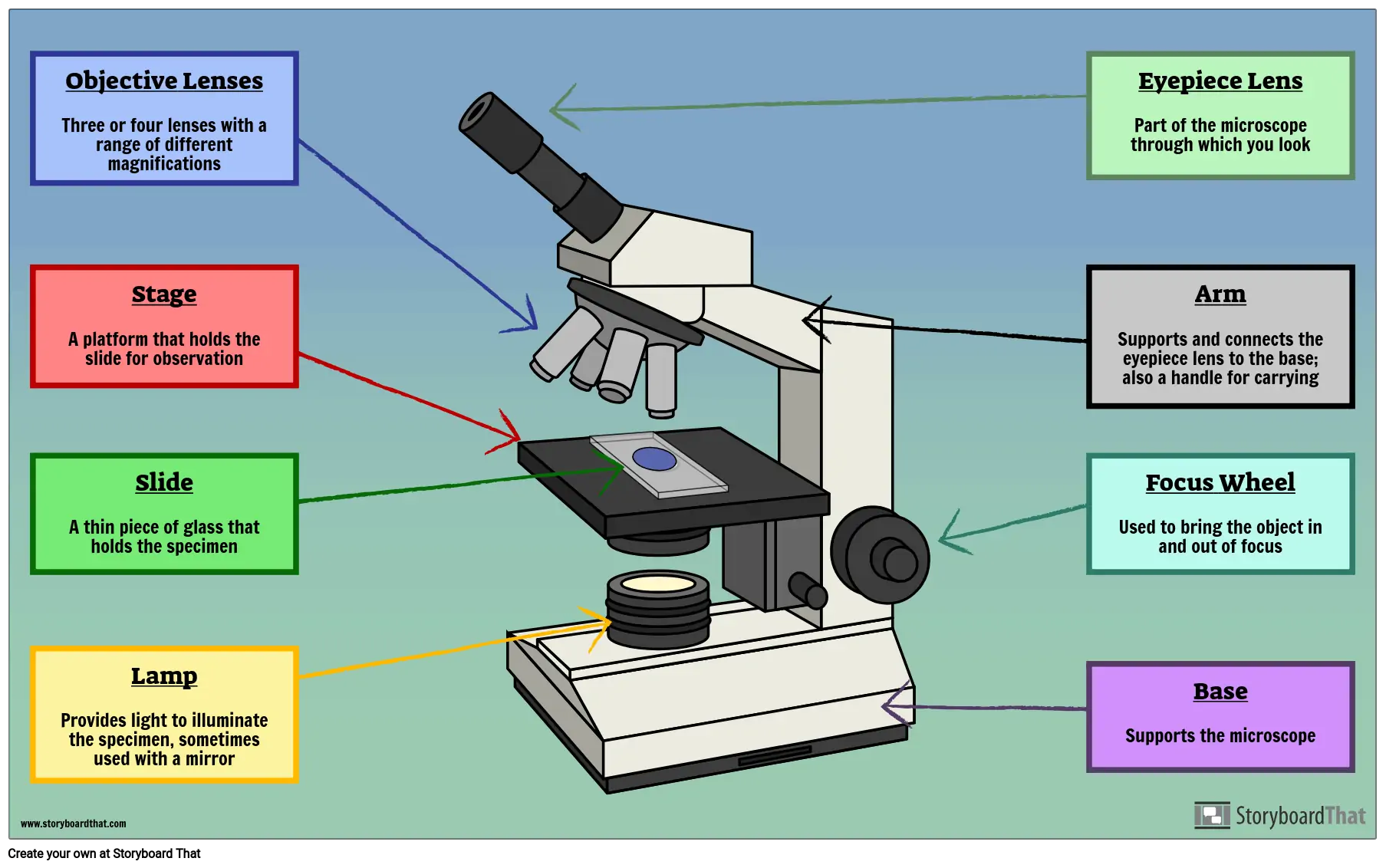
Parts of a Microscope Labeling Activity

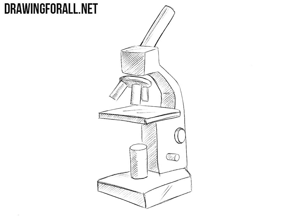
How to Draw a Microscope - Really Easy Drawing Tutorial
B1 E) Microscopy: Using Microscopes – AQA Combined Science ...
Label a microscope - Teaching resources
PRACTICAL BOOKLET - BIOLOGY4ISC

How to draw and label the parts of a microscope? What are at ...

Drawing Basics : How to Draw a Microscope | Drawings, Draw, Microscope

Anatomy of a Microscope - ppt download

Microscope Diagram Labeled, Unlabeled and Blank | Parts of a ...
Parts and Functions of a Compound Microscope Light
Free Microscope Drawing, Download Free Microscope Drawing png ...
Microscope Diagram Parts - ClipArt Best
Solved 7. The Microscope 1. In a compound microscope: a. The ...

parts of microscope with diagram

Compound Microscope: Parts of Compound Microscope

how to draw microscope step by step slow and medium speed
Black Microscope Icon On A Light Gray Background. Label Drawn ...

Label the microscope — Science Learning Hub

Microscope Parts worksheet
Post a Comment for "38 draw and label the microscope"