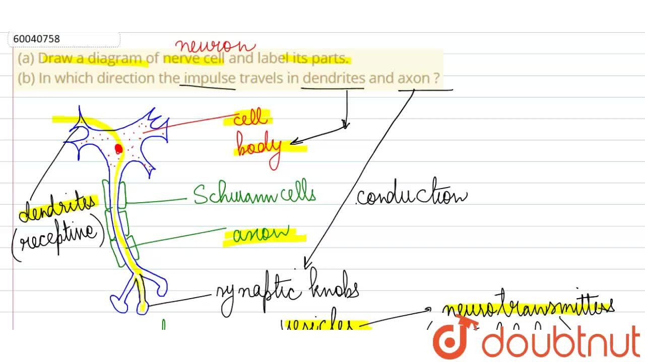
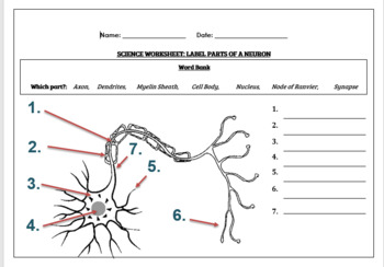
40 drawing of neuron and label of its parts
› science › in-in-class-11Synapse structure (video) | Khan Academy Let me start by drawing a big axon terminal, so I'll just blow up that axon terminal and make it really big here in green, and then I'll just draw a target type of shape to represent the target cell, which again could be another neuron, could be a muscle cell, or it could be a gland cell. Animal Cell Anatomy & Diagram - Enchanted Learning A small body located near the nucleus - it has a dense center and radiating tubules. The centrosomes is where microtubules are made. During cell division (mitosis), the centrosome divides and the two parts move to opposite sides of the dividing cell. The centriole is the dense center of the centrosome. Cytoplasm
Single Layer Perceptron in TensorFlow - Javatpoint Frank Rosenblatt first proposed in 1958 is a simple neuron which is used to classify its input into one or two categories. Perceptron is a linear classifier, and is used in supervised learning. It helps to organize the given input data. A perceptron is a neural network unit that does a precise computation to detect features in the input data. Perceptron is mainly used to classify the data …
Drawing of neuron and label of its parts
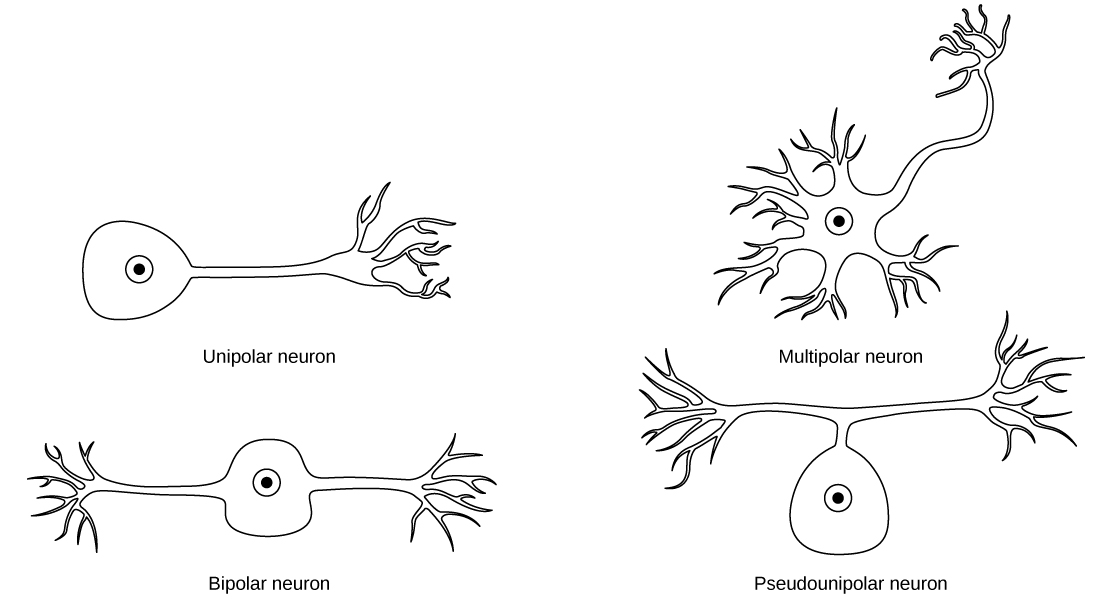
vas3k.com › blog › machine_learningMachine Learning for Everyone :: In simple words. With real ... Nov 21, 2018 · Neuron is a function with a bunch of inputs and one output. Its task is to take all numbers from its input, perform a function on them and send the result to the output. Here is an example of a simple but useful in real life neuron: sum up all numbers from the inputs and if that sum is bigger than N — give 1 as a result. Otherwise — zero. Types of Neurons: Parts, Structure, and Function - Verywell Health Summary. Neurons are responsible for transmitting signals throughout the body, a process that allows us to move and exist in the world around us. Different types of neurons include sensory, motor, and interneurons, as well as structurally-based neurons, which include unipolar, multipolar, bipolar, and pseudo-unipolar neurons. › doi › 10Cell-type profiling in salamanders identifies innovations in ... Sep 01, 2022 · After the divergence of mammals and sauropsids (reptiles and birds) ~320 million years ago, innovations in the pallium (i.e., the dorsal telencephalon) paved the way for advanced cognition. In mammals, the neocortex, with its characteristic six layers, evolved from a simpler ancestral cortex located in the dorsal pallium .
Drawing of neuron and label of its parts. PDF Worksheet for classes 1 and 2 - University of Minnesota Draw a generic neuron. Label its main parts. On your drawing of a neuron, indicate the direction of flow of information. Distinguish between the central and peripheral nervous systems. Draw a simple diagram of the lateral surface of a human brain. Draw the central and lateral sulci. Label the major brain regions visible in this view. To draw: A typical neuron and label it and give the function of each of ... Textbook solution for Biology (MindTap Course List) 11th Edition Eldra Solomon Chapter 41.2 Problem 2LO. We have step-by-step solutions for your textbooks written by Bartleby experts! Unit+3+Control+and+Coordination+Study+Guide.docx - Unit 3... Unit 3 Control and Coordination Study Guide Draw a picture of a neuron and label its key parts. ⮚ Dendrites go to soma (cell body)to the axon through myelin sheath and to the axon terminal. No synapse because we need a receptor to another neuron. If it has synapse it is between neurons. Cell-type profiling in salamanders identifies innovations in … 01/09/2022 · After the divergence of mammals and sauropsids (reptiles and birds) ~320 million years ago, innovations in the pallium (i.e., the dorsal telencephalon) paved the way for advanced cognition. In mammals, the neocortex, with its characteristic six layers, evolved from a simpler ancestral cortex located in the dorsal pallium .
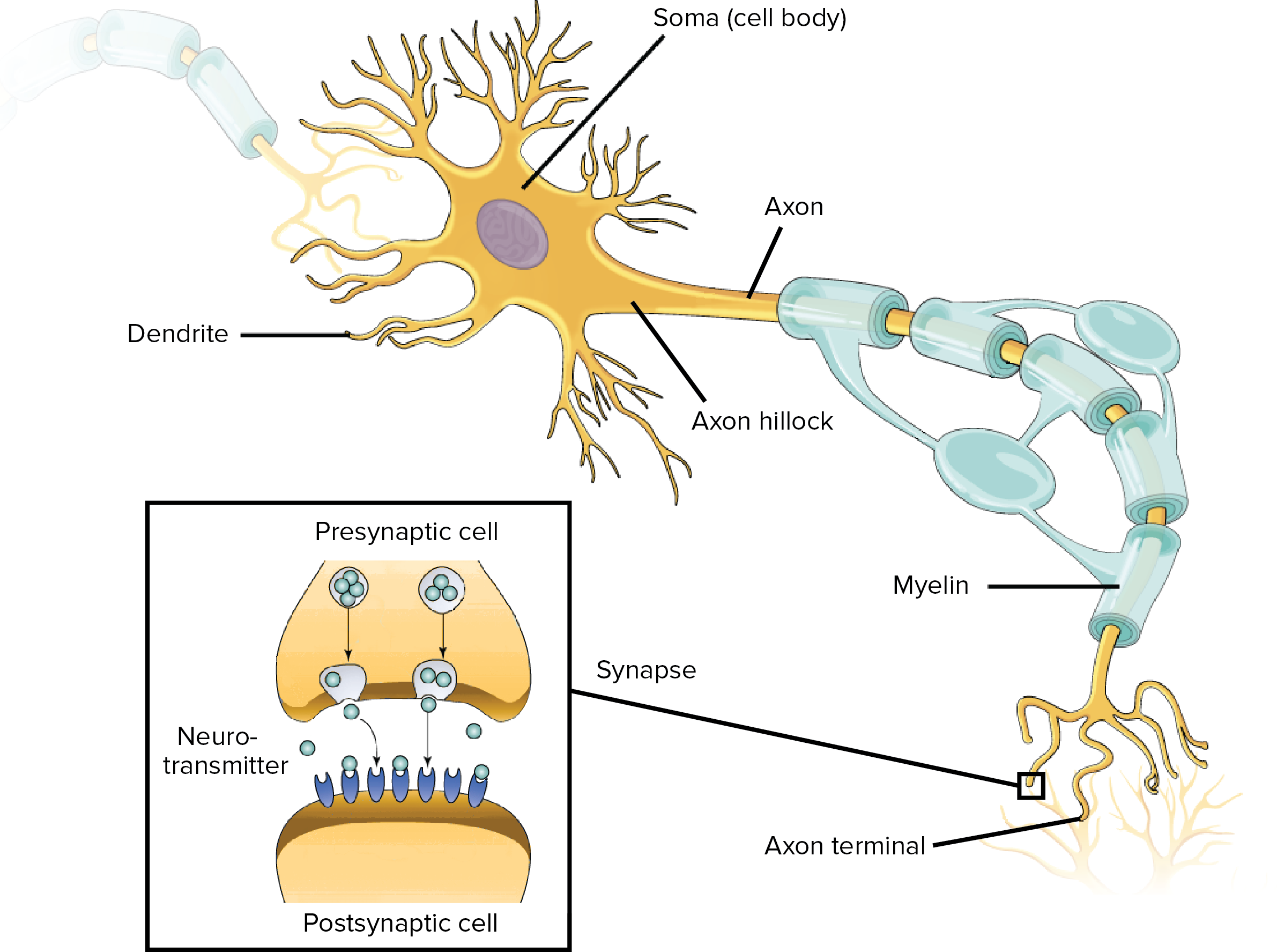
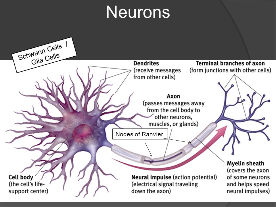
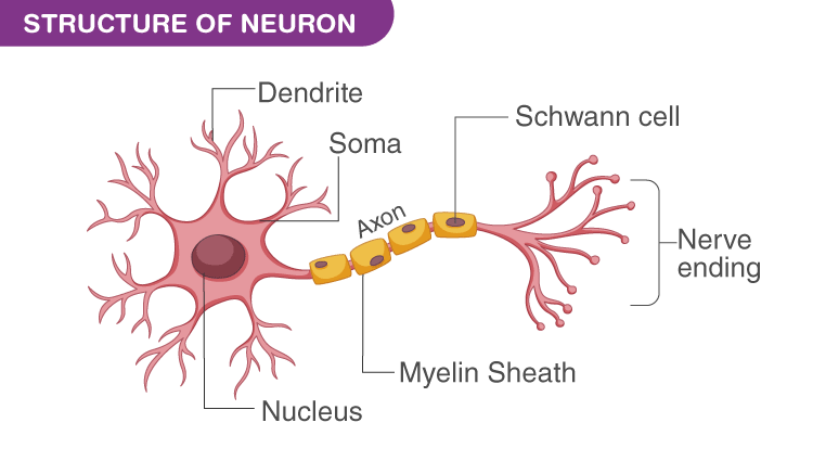
› Subjects › animalsAnimal Cell Anatomy & Diagram - Enchanted Learning A small body located near the nucleus - it has a dense center and radiating tubules. The centrosomes is where microtubules are made. During cell division (mitosis), the centrosome divides and the two parts move to opposite sides of the dividing cell. The centriole is the dense center of the centrosome. Cytoplasm What Is a Neuron? Diagrams, Types, Function, and More - Healthline Parts of a neuron Neurons vary in size, shape, and structure depending on their role and location. However, nearly all neurons have three essential parts: a cell body, an axon, and dendrites. Neuron Diagram || Diagram Of A Neuron || How To Draw A Neuron ... - YouTube Hello Everyone.Neuron Diagram || Diagram Of A Neuron || How To Draw A Neuron Step By Step For BeginnersNeuron Diagram, Diagram Of A Neuron, How To Draw A Neu... Parts of a Neuron and How Signals are Transmitted - Verywell Mind Axon. The axon is the elongated fiber that extends from the cell body to the terminal endings and transmits the neural signal. The larger the diameter of the axon, the faster it transmits information. Some axons are covered with a fatty substance called myelin that acts as an insulator.
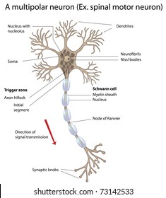
› single-layer-perceptron-inSingle Layer Perceptron in TensorFlow - Javatpoint A perceptron is a neural network unit that does a precise computation to detect features in the input data. Perceptron is mainly used to classify the data into two parts. Therefore, it is also known as Linear Binary Classifier. Perceptron uses the step function that returns +1 if the weighted sum of its input 0 and -1. Cell (biology) - Wikipedia Cell shapes. Cell shape, also called cell morphology, has been hypothesized to form from the arrangement and movement of the cytoskeleton. Many advancements in the study of cell morphology come from studying simple bacteria such as Staphylococcus aureus, E. coli, and B. subtilis. Different cell shapes have been found and described, but how and why cells form … Neurons (With Diagram) - Biology Discussion A neuron is a structural and functional unit of the neural tissue and hence the neural system. Certain neurons may almost equal the length of body itself. Thus neurons with longer processes (projections) are the longest cells in the body. Human neural system has about 100 billion neurons. Majority of the neurons occur in the brain. Structure of Neurons: What Is a Neuron? Types, Structure, Parts Structure of Neuron. Each neuron has a cell body, which is the central area of the neuron. It contains the nucleus and other structures common to all cells in the body, such as mitochondria. Neurons have highly branched fibres that reach out from the neuron are called dendritic trees. Each branch is called a dendrite.
a) Draw the structure of neuron and label cell body and axon ...
Diagram Quiz on Neuron Structure and Function (Labeling Quiz) 1. Identify the cell type in the above figure Liver Cell Cardiac Cell Nerve cell Skin cell 2. In the figure, labeled '1' receives impulses from adjacent neuron. It is called the Dendron Dendrite Axon Axonite 3. In the figure, labeled '2' is the short filaments from the cell body that carries impulses from dendrites to the cell body which is the
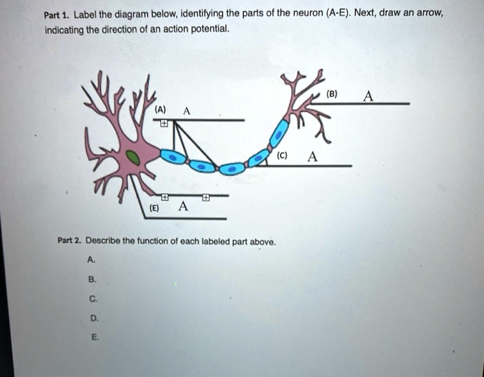
SOLVED: Part 1. Label the diagram below; identifying the ...
Label Parts of a Neuron Diagram | Quizlet Dendrites. receives impulses from other nerve cells. axon hillock. The cell body...the part of the cell that houses the nucleus and keeps the entire cell alive and functioning. Myelin Sheath. Surrounds the axon an insulates it from surrounding cells and tissues and making signal transitions faster and more efficient. Terminal Buttons.

Lesson Worksheet:Neurons | Nagwa
Answered: Label : lumen , globet cells , simply… | bartleby Solution for Label : lumen , globet cells , simply columnar epithelium , lamina propria , muscularis muscosa , submucosa . Skip to main content. close. Start your trial now! First week only $4.99! arrow_forward. Literature guides Concept explainers Writing guide Popular textbooks Popular high school textbooks Popular Q&A Business Accounting Economics Finance Leadership …
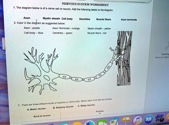
SOLVED: NERVOUS SYSTEM WORKSHEET nenve cell neuron. Add Ine ...
how to draw structure of neuron/neuron diagram labelled/diagram of ... Three Types Of Neurons Chemical Synapse Enteric Nervous System Glial Cells Psicologia The nervous system works through an interconnected network of billions of neurons. These neurons transmit information in the form of nerve impulses, across the nervous system and thus, coordinate the various functions of the body. X Xesus perez otero Psicologia

Nervous System
Draw a labelled diagram of neuron and label any four different parts ... Draw a labelled diagram of neuron and label any four different parts explain Here is the description of human neuron along with the diagram of the neuron and their parts. The neuron is a specialized and individual cell, which is also known as the nerve cell. A group of neurons forms a nerve. 1.
Overview of neuron structure and function (article) | Khan ...
Draw the diagram of neuron and label any two parts. - Toppr Ask Name the parts labelled a,b,c,d,e,f,g and h in the neuron drawn below. Medium. View solution. >. Draw a diagram of the human nerve cell. Justify its shape with regards to its function. Medium. View solution. >.

Draw a labelled diagram of a neuron with a myelin sheath.
Solved 4 Draw a neuron, label its parts, and describe their - Chegg Expert Answer. The brain is made up of billions of neurons. Neurons are specialized cells of the nervous system, which transmit signals all over the body. A neuron has these basic structures: the dendrites, the cell body, the axon …. View the full answer. Transcribed image text: 4 Draw a neuron, label its parts, and describe their functions.

What Is a Neuron? Diagrams, Types, Function, and More
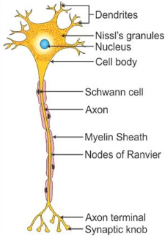
Draw a labelled diagram of the neuron and describe the structure of ... It consists of three major parts namely, Cell body, dendrites, Axon. Cell Body: It is irregular in shape or polyhedral. The cell body contains cytoplasm and certain granular bodies called Nissles granules which contain a group of ribosomes for protein synthesis.
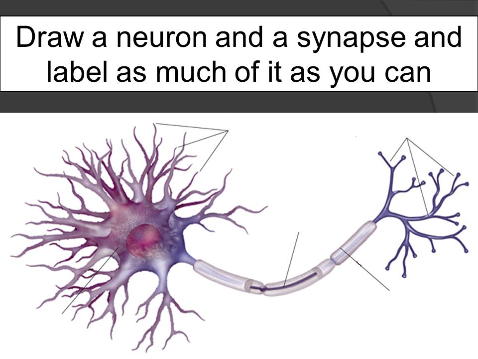
Draw a neuron and a synapse and label as much of it as you can
Draw diagram of a neuron and label its various parts. - Brainly Draw diagram of a neuron and label its various parts. - 42449081 SnehaRawat9898 SnehaRawat9898 4 weeks ago Biology Secondary School answered Draw diagram of a neuron and label its various parts. 2 See answers ...
Draw a neuron and a synapse and label as much of it as you ...
The 9 parts of a neuron (and their functions) - medical - 2022 A neuron is a type of cell. Just like those that make up our muscles, liver, heart, skin, etc. But the key point is that each type of cell adapts both its morphology and structure depending on what function they have to perform. Y neurons have a very different purpose than other cells in the body.
Neuron Cell Worksheets - Superstar Worksheets
medical-dictionary.thefreedictionary.com › painPain | definition of pain by Medical dictionary The discomfort signals actual or potential injury to the body. However, pain is more than a sensation, or the physical awareness of pain; it also includes perception, the subjective interpretation of the discomfort. Perception gives information on the pain's location, intensity, and something about its nature.
Solved] Draw a unipolar, bipolar and multipolar neuropn and ...
Machine Learning for Everyone - vas3k.com 21/11/2018 · Machine Learning is like sex in high school. Everyone is talking about it, a few know what to do, and only your teacher is doing it. If you ever tried to read articles about machine learning on the Internet, most likely you stumbled upon two types of them: thick academic trilogies filled with theorems (I couldn’t even get through half of one) or fishy fairytales about …

Label Parts of a Neuron Diagram | Quizlet
Physiology exam2 learning outcomes.docx - Physiology Exam 2... View Physiology exam2 learning outcomes.docx from BIOL 2213 at University of Arkansas. Physiology Exam 2 Chapter 7 - Learning Outcomes From the Reading: - Draw a neuron, label its parts, and describe
Draw the structure of a neuron and explain its function ...
Neurons: Structure and Functions (With Diagram) - Psychology Discussion It is the coordinating neuron. The neurons are of three kinds according to their functions. The sensory neurons conduct nerve currents from the sense-organs to the sensory centres. The motor neurons which terminate in muscles carry nerve currents from the motor centres to the muscles. The central neurons connect sensory neurons with motor neurons.

Senses 5
Labeled Neuron Diagram | Science Trends Neurons are a type of cell and are the fundamental constituents of the nervous system and brain. Neurons take in stimuli and convert them to electrical and chemical signals that are sent to our brain. There are 3 major kinds of neurons in the spinal cord: sensory, motor, and interneurons.

Label: Parts of a Common Neuron Flashcards | Quizlet
A Guide to Understand Neuron with Neuron Diagram The students can find the neuron labeled diagram in this option. Step 3: Once the students have selected their diagrams, they can smoothly work on them. They can modify the diagram according to their choice. It can help them to get a high-quality neuron labeled diagram apt for their projects and dissertation papers.

Labeled Neuron Diagram | Science Trends
Synapse structure (video) | Khan Academy Let me start by drawing a big axon terminal, so I'll just blow up that axon terminal and make it really big here in green, and then I'll just draw a target type of shape to represent the target cell, which again could be another neuron, could be a muscle cell, or it could be a gland cell. And then in the central nervous system, covering most of the synapses are the end feet of astrocytes. …
16.1 Neurons and Glial Cells – Concepts of Biology – 1st ...
AP Psychology ALL Terms Flashcards | Quizlet the junction between the axon tip of the sending neuron and the dendrite or cell body of the receiving neuron. The tiny gap at this junction is called the synaptic gap or cleft . Neurotransmitters. chemical messengers that traverse the synaptic gaps between neurons. When released by the sending neuron, neurotransmitters travel across the synapse and bind to …
Draw a labelled diagram of a neuron - CBSE Class 11 - Learn ...
Nervous System - Label the Neuron - TheInspiredInstructor.com Choose the correct names for the parts of the neuron. (1) (2) (3) (4) (5) (6) This neuron part receives messages from other neurons. (7) This neuron part sends on messages to other neurons. (8) This neuron part gives messages to muscle tissue. (9) This neuron part processes incoming messages.
What are the parts of the neuron and their function? - Quora
A Labelled Diagram of Neuron with Detailed decription - Collegedunia A neuron is a type of cell that is largely responsible for conveying information via electrical and chemical impulses. The brain, spinal cord, and peripheral nerves all contain them. The nerve cell is another name for a neuron. The structure of a neuron changes depending on its form and size, as well as its function and location.

Q3 Given below is the diagram of a neuron Name the parts ...
Anatomy and Physiology – Easy Peasy All-in-One High School Wow, that’s a lot but that’s only the beginning! Each part of the cell has a specific structure AND function. This course will explore the anatomy, the structure of the human body and its parts, and the physiology or the functions of the human body and its parts. Human Anatomy and Physiology is where biology and chemistry merge to create ...

Neuron - Wikipedia
Neuron Diagram & Types | Ask A Biologist - Arizona State University Multipolar neurons have one axon and many dendritic branches. These carry signals from the central nervous system to other parts of your body such as your muscles and glands. Unipolar neurons are also known as sensory neurons. They have one axon and one dendrite branching off in opposite directions from the cell body.
Neuron Diagram Teaching Resources | Teachers Pay Teachers
Draw a Labeled Diagram of a Myelinated Neuron. - Biology Draw a Labeled Diagram of a Myelinated Neuron. CISCE ICSE Class 10. Question Papers 359. Textbook Solutions 25661. MCQ Online Tests 6. Important Solutions 3381. Question Bank Solutions 24997. Concept Notes & Videos 619. ... Sketch and Label the Diagram | Q 3. Advertisement Remove all ads.

Polarity of Cell Membranes - ScienceDirect
› questions-and-answers › labelAnswered: Label : lumen , globet cells , simply… | bartleby Solution for Label : lumen , globet cells , simply columnar epithelium , lamina propria , muscularis muscosa , submucosa

12.143 Akson Gambar, Foto Stok & Vektor | Shutterstock
Answered: Draw a neuron, label its parts, and… | bartleby Relate the structure of the neuron to its function. arrow_forward Diagram and label the basic features of a neuron,indicating where information is received and where it is sent. arrow_forward Draw an arrow showing the direction that an impulse travels along a neuron arrow_forward Describe the anatomy of a neuron. arrow_forward

Well labelled diagram of neuron preferably hand drawn ...
Pain | definition of pain by Medical dictionary Pain Definition Pain is an unpleasant feeling that is conveyed to the brain by sensory neurons. The discomfort signals actual or potential injury to the body. However, pain is more than a sensation, or the physical awareness of pain; it also includes perception, the subjective interpretation of the discomfort. Perception gives information on the pain's ...

6.5.2 Draw and label a diagram of the structure of a motor neuron
A Labelled Diagram Of Neuron with Detailed Explanations - BYJUS Diagram Of Neuron with Labels Here is the description of human neuron along with the diagram of the neuron and their parts. The neuron is a specialized and individual cell, which is also known as the nerve cell. A group of neurons forms a nerve.
Motor Neuron Detailed Accurate Labeled Stock Vector (Royalty ...
› doi › 10Cell-type profiling in salamanders identifies innovations in ... Sep 01, 2022 · After the divergence of mammals and sauropsids (reptiles and birds) ~320 million years ago, innovations in the pallium (i.e., the dorsal telencephalon) paved the way for advanced cognition. In mammals, the neocortex, with its characteristic six layers, evolved from a simpler ancestral cortex located in the dorsal pallium .

Label the parts of a neuron in Figure 7.5. - Sarthaks ...
Types of Neurons: Parts, Structure, and Function - Verywell Health Summary. Neurons are responsible for transmitting signals throughout the body, a process that allows us to move and exist in the world around us. Different types of neurons include sensory, motor, and interneurons, as well as structurally-based neurons, which include unipolar, multipolar, bipolar, and pseudo-unipolar neurons.

How to Draw a Neuron and Label Its Parts
vas3k.com › blog › machine_learningMachine Learning for Everyone :: In simple words. With real ... Nov 21, 2018 · Neuron is a function with a bunch of inputs and one output. Its task is to take all numbers from its input, perform a function on them and send the result to the output. Here is an example of a simple but useful in real life neuron: sum up all numbers from the inputs and if that sum is bigger than N — give 1 as a result. Otherwise — zero.

Neuron Illustrations | Casey Henley, PhD

n Illustration of neurotransmitters being released from the ...
a) Draw a diagram of nerve cell and label its parts. (b) In ...

Dendrite - Wikipedia

draw a labelled diagram of a neuron j8t76t088 -Biology ...

Neurons worksheet

Histology of the Peripheral Nerves and Light Microscopy ...
Draw the structure of a neuron and label and following parts ...

What Is a Neuron? Diagrams, Types, Function, and More
Neurons Worksheet Teaching Resources | Teachers Pay Teachers
Post a Comment for "40 drawing of neuron and label of its parts"