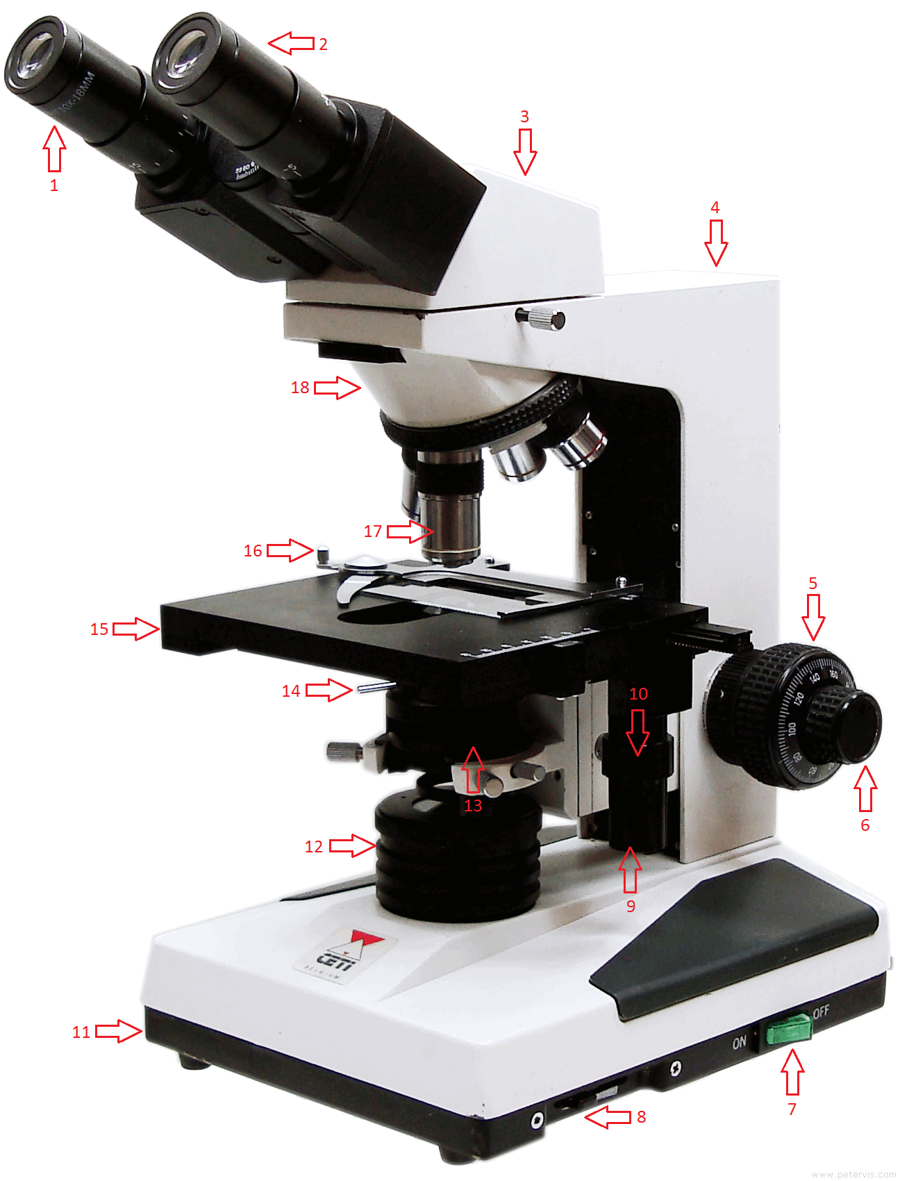
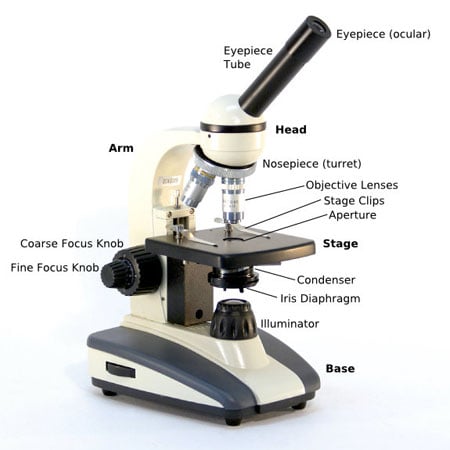
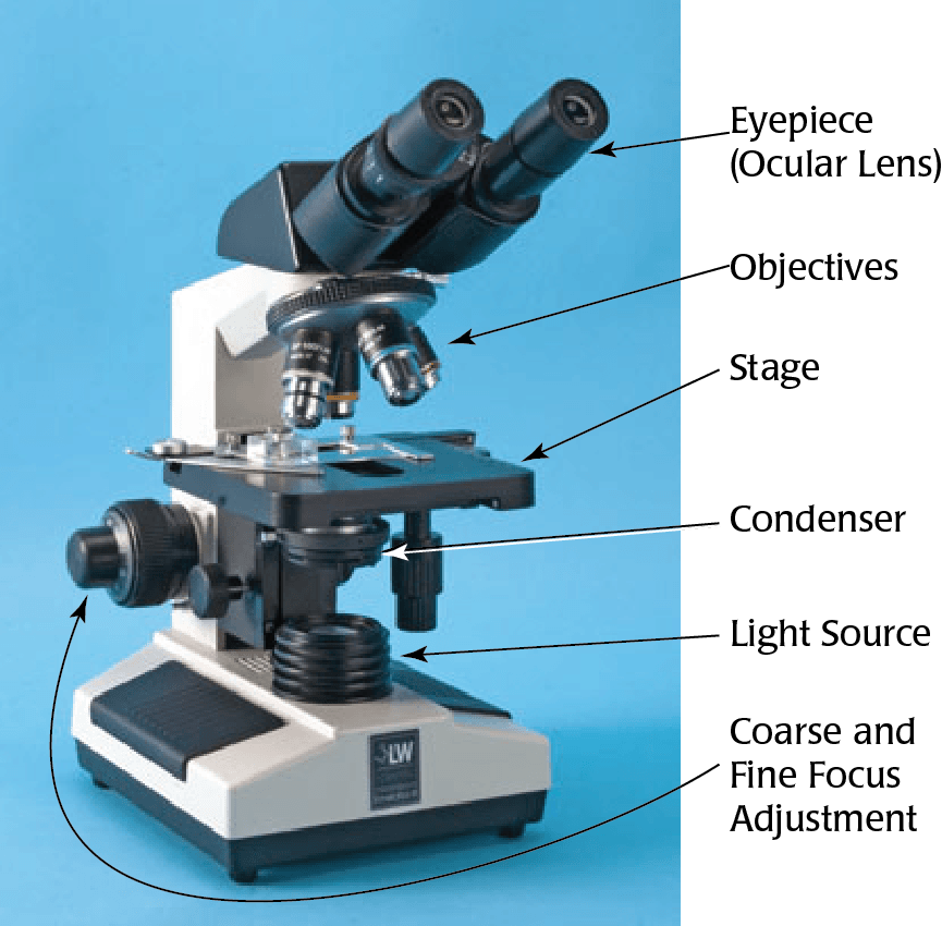
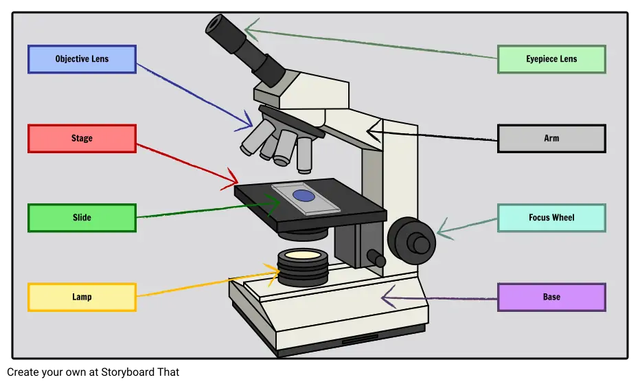
45 microscope labeled
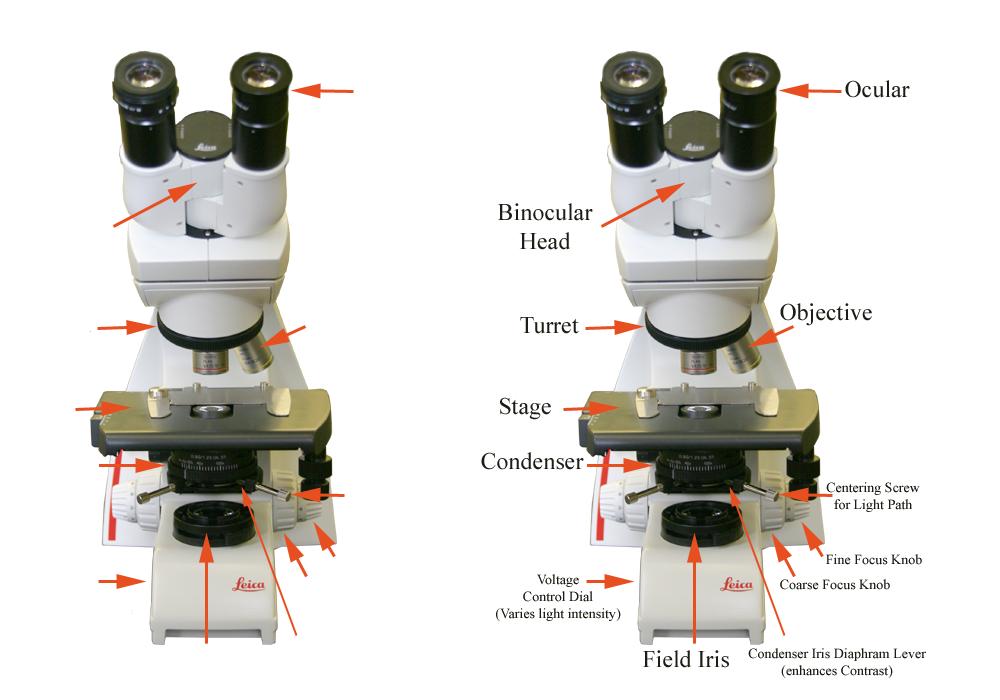
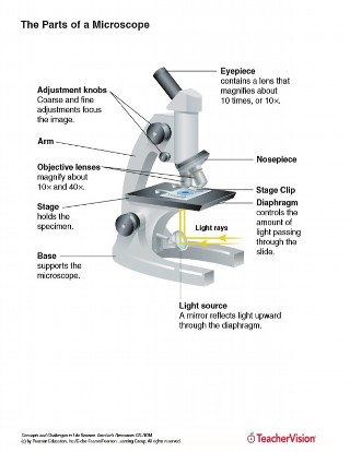
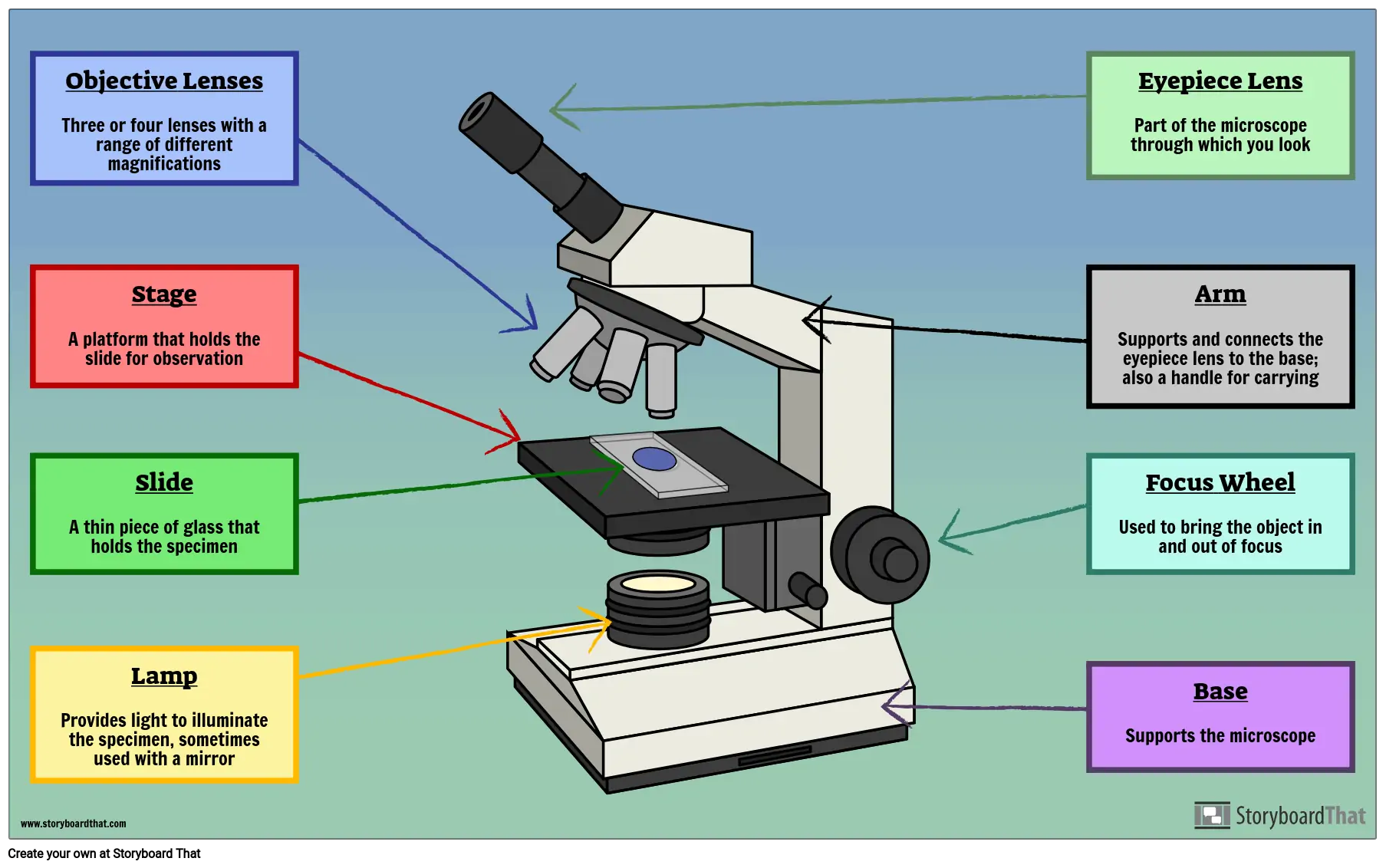
Microscope Parts, Function, & Labeled Diagram - slidingmotion Microscope Parts Labeled Diagram The principle of the Microscope gives you an exact reason to use it. It works on the 3 principles. Magnification Resolving Power Numerical Aperture. Parts of Microscope Head Base Arm Eyepiece Lens Eyepiece Tube Objective Lenses Nose Piece Adjustment Knobs Stage Aperture Microscopic Illuminator Condenser Lens Labeling the Parts of the Microscope | Microscope World Resources Labeling the Parts of the Microscope This activity has been designed for use in homes and schools. Each microscope layout (both blank and the version with answers) are available as PDF downloads. You can view a more in-depth review of each part of the microscope here. Download the Label the Parts of the Microscope PDF printable version here.
Observing Onion Cells Under The Microscope » Microscope Club Observing onion cells under a microscope is a fun and easy activity for students and hobbyists alike. Onion epidermal cells appear as a single thin layer and look highly organized and structured in terms of shape and size. Certain parts of the cell are also clearly distinguishable with or without staining, making the activity even easier and ...

Microscope labeled
laboratoryinfo.com › labeling-parts-of-the-microscopeParts of the Microscope with Labeling (also Free Printouts) Mar 07, 2022 · Parts of the Microscope with Labeling (also Free Printouts) By Editorial Team March 7, 2022 A microscope is one of the invaluable tools in the laboratory setting. It is used to observe things that cannot be seen by the naked eye. Table of Contents 1. Eyepiece 2. Body tube/Head 3. Turret/Nose piece 4. Objective lenses 5. Knobs (fine and coarse) 6. Simple Squamous Epithelium under a Microscope with a Labeled Diagram ... Histological features of lung parenchyma with microscopic slide images and labeled diagrams. The lung's alveoli give the honeycomb appearance in the parenchyma and lines by flattened simple squamous epithelium. These alveoli are thin-walled and fills with air. From the lung parenchyma labeled diagram, you might identify the following ... Virtual Microscope | NCBioNetwork.org Learning Objectives. Upon completion of this exercise, you will be able to: List the basic components of a typical microscope. Describe the use of lens power and eyepiece powers. Calculate the magnification of a microscope based on the selected lens. Discuss the care of an use of a typical microscope.
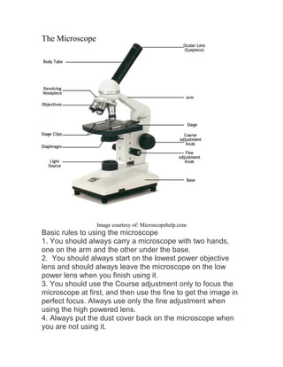
Microscope labeled. Parts of a Microscope Labeling Activity - Storyboard That In order to study cells in detail, it's important that students learn how to use a microscope. Before using microscopes in the classroom, they should understand the names and function of each part. In this activity, students will create a poster of a microscope with labeled parts. Students will identify and describe the microscope parts and ... rsscience.com › compound-microscope-parts-labeledCompound Microscope Parts – Labeled Diagram and their Functions The term “compound” refers to the microscope having more than one lens. Basically, compound microscopes generate magnified images through an aligned pair of the objective lens and the ocular lens. In contrast, “simple microscopes” have only one convex lens and function more like glass magnifiers. [In this figure] Two “antique ... Parts of a microscope with functions and labeled diagram - Microbe Notes Microscopes are instruments that are used in science laboratories to visualize very minute objects such as cells, and microorganisms, giving a contrasting image that is magnified. Microscopes are made up of lenses for magnification, each with its own magnification powers. Microscope Labeling Game - PurposeGames.com An unregistered player played the game 32 minutes ago About this Quiz This is an online quiz called Microscope Labeling Game There is a printable worksheet available for download here so you can take the quiz with pen and paper. This quiz has tags. Click on the tags below to find other quizzes on the same subject. Science microsope
Parts of Stereo Microscope (Dissecting microscope) - labeled diagram ... Major microscope brands (Zeiss, Olympus, Nikon, Amscope, Omano, Leica …) all produce stereomicroscopes. Photo source: AOMEKIE, Bresser, Swift The name "stereo" comes from the term "stereoscopic," meaning, viewing by two different angles to create an impression of depth and solidity. bone under microscope labeled Bone microscopic structure clipart etc usf micro edu tiff medium resolution. bone under microscope labeled. Microscopic Structure of Bone | ClipArt ETC. 11 Pics about Microscopic Structure of Bone | ClipArt ETC : Last week's anatomic structure was a microscopic view of an osteon. The, Skeletal System - Dr. Hunter's Anatomy and Physiology and ... Fluorescence Microscopy - Explanation and Labelled Images Another use of fluorescence imaging is Fluorescence Speckle Microscopy. It is a technology that uses fluorescence labeled macromolecular assemblies such as cytoskeletal protein to study movement and turnover rates. Fluorescence microscopy staining also is helpful in the field of mineralogical applications. It is routinely used for the study of ... Microscope Labeling - The Biology Corner The labeling worksheet could be used as a quiz or as part of direct instruction where students label the microscope as you go over what each part is used for. The google slides shown below have the same microscope image with the labels for students to copy.
Microscope Types (with labeled diagrams) and Functions Simple microscope labeled diagram Simple microscope functions It is used in industrial applications like: Watchmakers to assemble watches Cloth industry to count the number of threads or fibers in a cloth Jewelers to examine the finer parts of jewelry Miniature artists to examine and build their work Also used to inspect finer details on products Label the microscope — Science Learning Hub All microscopes share features in common. In this interactive, you can label the different parts of a microscope. Use this with the Microscope parts activity to help students identify and label the main parts of a microscope and then describe their functions. Drag and drop the text labels onto the microscope diagram. › photos › microscope-labeledMicroscope Labeled Pictures, Images and Stock Photos Browse 49 microscope labeled stock photos and images available, or start a new search to explore more stock photos and images. Newest results. Fluorescent Imaging immunofluorescence of cancer cells growing in 2D with nuclei in blue, cytoplasm in red and DNA damage foci in green Fluorescent Imaging immunofluorescence of cancer cells growing in ... Microscope Parts & Functions - AmScope Invented by a Dutch spectacle maker in the late 16th century, compound light microscopes use two sets of lenses to magnify images for study and observation. The first set of lenses are the oculars, or eyepieces, that the viewer looks into; the second set of lenses are the objectives, which are closest to the specimen.
› parts-of-a-compound-microscopeMicroscope Parts and Functions Microscope Parts and Functions With Labeled Diagram and Functions How does a Compound Microscope Work?. Before exploring microscope parts and functions, you should probably understand that the compound light microscope is more complicated than just a microscope with more than one lens.. First, the purpose of a microscope is to magnify a small object or to magnify the fine details of a larger ...
A Study of the Microscope and its Functions With a Labeled Diagram ... To better understand the structure and function of a microscope, we need to take a look at the labeled microscope diagrams of the compound and electron microscope. These diagrams clearly explain the functioning of the microscopes along with their respective parts. Man's curiosity has led to great inventions. The microscope is one of them.
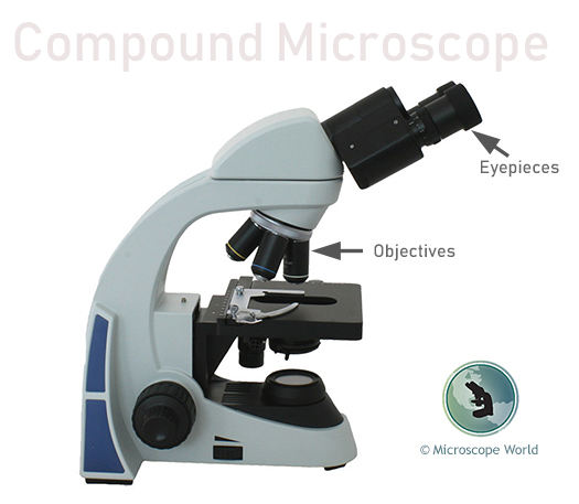
Compound Microscope Parts, Functions, and Labeled Diagram Common compound microscope parts include: Eyepiece (ocular lens) with or without Pointer: The part that is looked through at the top of the compound microscope. Eyepieces typically have a magnification between 5x & 30x. Monocular or Binocular Head: Structural support that holds & connects the eyepieces to the objective lenses.
Compound Microscope: Definition, Diagram, Parts, Uses, Working ... - BYJUS Compound microscope is a type of optical microscope that is used for obtaining a high-resolution image. There are more than two lenses in a compound microscope. Learn about the working principle, parts and uses of a compound microscope along with a labeled diagram here.
Parts of the Microscope quiz | Science Quiz - Quizizz 10 Questions Show answers. Question 1. SURVEY. 30 seconds. Q. Label the part of the microscope. What is part A? answer choices. eyepiece.
Light Microscope- Definition, Principle, Types, Parts, Labeled Diagram ... A light microscope is a biology laboratory instrument or tool, that uses visible light to detect and magnify very small objects and enlarge them. They use lenses to focus light on the specimen, magnifying it thus producing an image. The specimen is normally placed close to the microscopic lens.
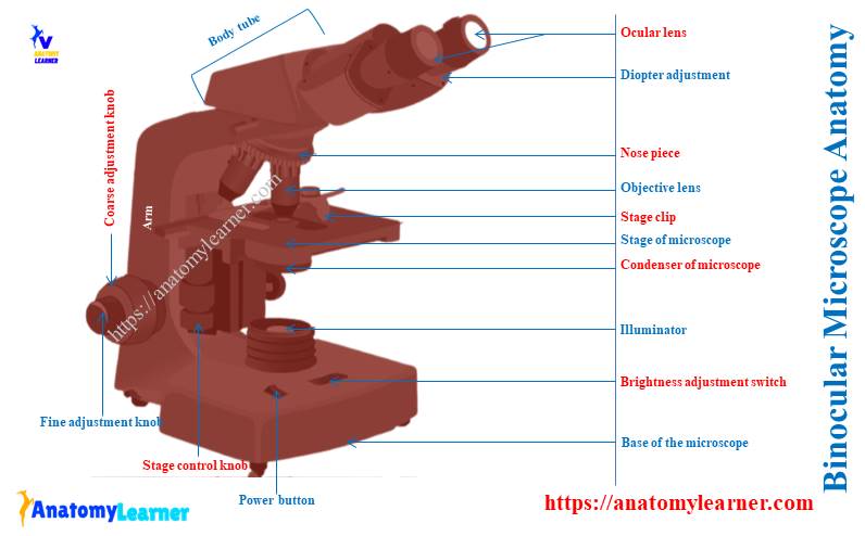
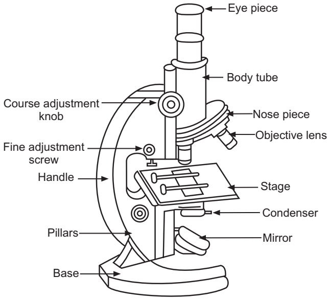
microscopespot.com › the-parts-of-a-microscopeParts of a Microscope – The Comprehensive Guide Microscope Parts Labeled: Parts of A Microscope 1. Eyepiece Lens and Eyepiece Tube 2. Objective Lens 3. Tube 4. Base 5. Arm 6. Illuminator 7. Stage or platform 8. Stage Clips 9. Rotating Turret or Nosepiece 10. Rack Stop 11. Condenser Lens 12. Iris or Diaphragm 13. Coarse Adjustment Knob 14. Fine Adjustment Knob 15. Power Switch
researchtweet.com › microscope-parts-labeled-diagram-and-functionsMicroscope, Microscope Parts, Labeled Diagram, and Functions Sep 03, 2022 · Revolving Nosepiece or Turret: Turret is the part of the microscope that holds two or multiple objective lenses and helps to rotate objective lenses and also helps to easily change power. Objective Lenses: Three are 3 or 4 objective lenses on a microscope. The objective lenses almost always consist of 4x, 10x, 40x and 100x powers. The most common eyepiece lens is 10x and when it coupled with ...
microscopewiki.com › simple-microscopeSimple Microscope - Diagram (Parts labelled), Principle, Formula... Feb 23, 2022 · Simple microscope is a magnification apparatus that uses a combination of double convex lens to form an enlarged, erect image of a specimen. The working principle of a simple microscope is that when a lens is held close to the eye, a virtual, magnified and erect image of a specimen is formed at the least possible distance from which a human eye ...
Observing Yeast Under The Microscope » Microscope Club Microscopy techniques Image sourced from researchgate.net. Yeast can be viewed under the microscope through two different microscopy techniques- bright field microscopy and fluorescence microscopy. For one thing, yeast and buds can be seen under a high magnification (1000x) bright field microscope, such as a compound microscope.
Binocular Microscope Anatomy - Parts and Functions with a Labeled ... Now, I will discuss the details anatomy of the light compound microscope with the labeled diagram. Why it is called binocular: because it has two ocular lenses or an eyepiece on the head that attaches to the objective lens, this ocular lens magnifies the image produced by the objective lens. Binocular microscope parts and functions
Light microscopes - Cell structure - Edexcel - BBC Bitesize The compound microscope uses two lenses to magnify the specimen: the eyepiece and an objective lens. In most microscopes, there is a choice of objectives to use. Magnification can therefore be ...
Virtual Microscope | NCBioNetwork.org Learning Objectives. Upon completion of this exercise, you will be able to: List the basic components of a typical microscope. Describe the use of lens power and eyepiece powers. Calculate the magnification of a microscope based on the selected lens. Discuss the care of an use of a typical microscope.
Simple Squamous Epithelium under a Microscope with a Labeled Diagram ... Histological features of lung parenchyma with microscopic slide images and labeled diagrams. The lung's alveoli give the honeycomb appearance in the parenchyma and lines by flattened simple squamous epithelium. These alveoli are thin-walled and fills with air. From the lung parenchyma labeled diagram, you might identify the following ...
laboratoryinfo.com › labeling-parts-of-the-microscopeParts of the Microscope with Labeling (also Free Printouts) Mar 07, 2022 · Parts of the Microscope with Labeling (also Free Printouts) By Editorial Team March 7, 2022 A microscope is one of the invaluable tools in the laboratory setting. It is used to observe things that cannot be seen by the naked eye. Table of Contents 1. Eyepiece 2. Body tube/Head 3. Turret/Nose piece 4. Objective lenses 5. Knobs (fine and coarse) 6.
















Post a Comment for "45 microscope labeled"