39 sarcomere structure labeled
Sarcomere Labeling Quiz - PurposeGames.com Sarcomere Labeling by emcanallen 52,724 plays 8 questions ~ 20 sec More 16 5.00 (you: not rated) Language English Tries Unlimited [?] Last Played February 22, 2022 - 12:00 am There is a printable worksheet available for download here so you can take the quiz with pen and paper. Remaining 0 Correct 0 Wrong 0 Press play! 0% 10:00.0 Highscores The Sarcomere and Sliding Filaments in Muscular Contraction: Definition ... A given myofibril contains approximately 10,000 sarcomeres, each of which is about 3 micrometers in length. While each sarcomere is small, several sarcomeres added together span the length of the ...
Page: Journal of Hand Surgery The Journal of Hand Surgery publishes original, peer-reviewed articles related to the pathophysiology, diagnosis, and treatment of diseases and conditions of the upper extremity; these include both clinical and basic science studies, along with case reports.
Sarcomere structure labeled
› books › NBK9926Glossary - The Cell - NCBI Bookshelf A sheetlike secondary structure of a polypeptide chain, formed by hydrogen bonding between amino acids located in different regions of the polypeptide. bacteriophage. A bacterial virus. basal body. A structure similar to a centriole that initiates the growth of axonemal microtubules and anchors cilia and flagella to the surface of the cell ... Labeled Sarcomere Diagram A sarcomere is the basic unit of striated muscle tissue. It is the repeating unit between two Z lines. Skeletal muscles are composed of tubular muscle cells which. Sarcomeres are composed of thick filaments and thin filaments. The thin filaments Look at the diagram above and realize what happens as a muscle contracts. Sarcomere Structure: Actin & Myosin - Notes - NinjaNerd Medicine Sarcomere Structure: Actin & Myosin. Musculoskeletal. . Physiology. Get access to all our resources including notes and illustrations when you sign up to become a Ninja Nerd member. Become a Member. . View Notes.
Sarcomere structure labeled. 10.2 Skeletal Muscle - Anatomy & Physiology The Sarcomere. A sarcomere is defined as the region of a myofibril contained between two cytoskeletal structures called Z-discs (also called Z-lines or Z-bands), and the striated appearance of skeletal muscle fibers is due to the arrangement of the thick and thin myofilaments within each sarcomere (Figure 10.2.2). › articles › s44161/022/00057-1Metabolic characterization of hypertrophic cardiomyopathy in ... May 09, 2022 · Top ten significantly increased or decreased metabolites of FC in each group are labeled. The horizontal line denotes FDR cutoff of 0.05 and the vertical lines denote a FC of 1.5 or 0.67. biologydictionary.net › sarcomereSarcomere - Definition, Structure, Function and Quiz ... Mar 28, 2019 · Sarcomere definition. A sarcomere is the functional unit of striated muscle. This means it is the most basic unit that makes up our skeletal muscle. Skeletal muscle is the muscle type that initiates all of our voluntary movement. Herein lies the sarcomere’s main purpose. Sarcomeres are able to initiate large, sweeping movement by contracting ... What is a Sarcomere? - Parts & Contraction - Study.com Each sarcomere has a central A-band which consists of thick filaments and two halves of I-band which has thin filaments. The I-bands from two neighboring sarcomeres meet at the Z-line, while the...
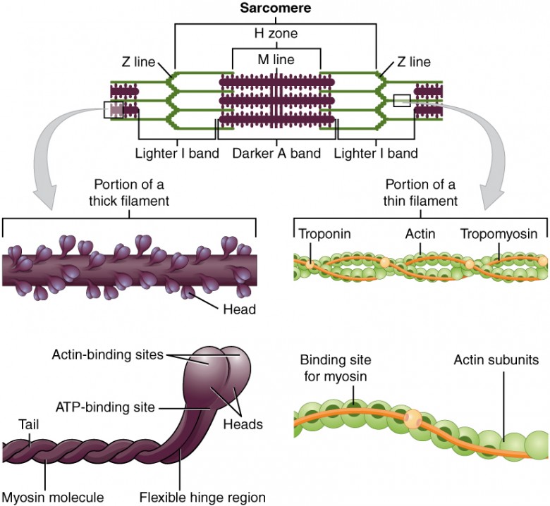
Describe the structure of sarcomere. - Toppr Ask Sarcomeres are composed of long, fibrous proteins as filaments that slide past each other when a muscle contracts or relaxes. Two of the important proteins are:- 1) Myosin forms the thick filament. Myosin has a long fibrous tail and a globular head, which binds to actin. Its head also binds to ATP, which is the source of energy for muscle movement. Sarcomere Structure : Mnemonic | Epomedicine Z is the final alphabet: Z lines represents the end of sarcomere M for middle: M line represents the midline of sarcomere I is a thin letter: I band has only thin filaments H is a thick letter: H zone has only thick filaments A is a hybrid of "I" and "H": A band has both thin and thick filaments (remains constant during contraction) Sarcomere: Structure and Parts, Functions and Histology Each sarcomere consists of thick, thin beams of the proteins mentioned above, which together are called myofilaments. By expanding a portion of the myofilaments, you can identify the molecules that make them up. The thick filaments are made of myosin, while the fine filaments are made of actin. Free ATI TEAS Science Diagnostic Test (50 Questions) - NurseHub WebThis free ATI TEAS 7 Science Practice Test will give you an accurate feel of the exam. There are 50 questions. You will receive a detailed score report at the end of the test, so we recommend you use this as a diagnostic test to see which topics or skills you should focus on the most.. Sign up for our Question of the Day email list to get even more free TEAS …
› topics › medicine-andSkeletal Muscle Cell - an overview | ScienceDirect Topics Skeletal Muscle Cell. In cultured skeletal muscle cells and C2C12 myotubes, Ucn 2 inhibits insulin-induced Akt and ERK1/2 phosphorylation, consistent with the hypothesis that Ucn 2 functions as a local negative regulator of glucose uptake in skeletal muscle and suggests the possibility that suppression of the Ucn 2/CRH-R2 pathway may provide benefits in insulin-resistant states such as type 2 ... Sarcomere : Anatomy of Muscle Structure A sarcomere is the basic functional within muscle cells. This unit is distinctive in some types of muscle tissue. It can be observed on microscope slides due to the striated nature of both skeletal muscle and cardiac muscle. (Although smooth muscle has a similar contractile mechanism, is not as highly organized and does not show striations). Sarcomere Labeling Diagram | Quizlet Sarcomere The smallest contractile unit of muscle; extends from one Z disc to the next H Band The band at the middle of the A Band, where only myosin is found A Band The darkest area that runs the length of the myosin, including where actin and myosin overlap I Band On either side of the A Band is the I band, where only the Actin is found Z Disk Sarcomere - Definition, Structure, Function and Quiz - Biology … Web28.03.2019 · They were able to visualize the physical lengthening of the sarcomere in its relaxed state, and the shortening in its contracted state. Their observations led to the discovery of sarcomere zones. The figure depicts the structure of a Sarcomere. (Each zone is labeled).
Draw the diagram of a sarcomere of skeletal muscle showing ...
Sarcomere - an overview | ScienceDirect Topics A sarcomere is the basic contractile unit of muscle fiber. Each sarcomere is composed of two main protein filaments—actin and myosin—which are the active structures responsible for muscular contraction. The most popular model that describes muscular contraction is called the sliding filament theory.
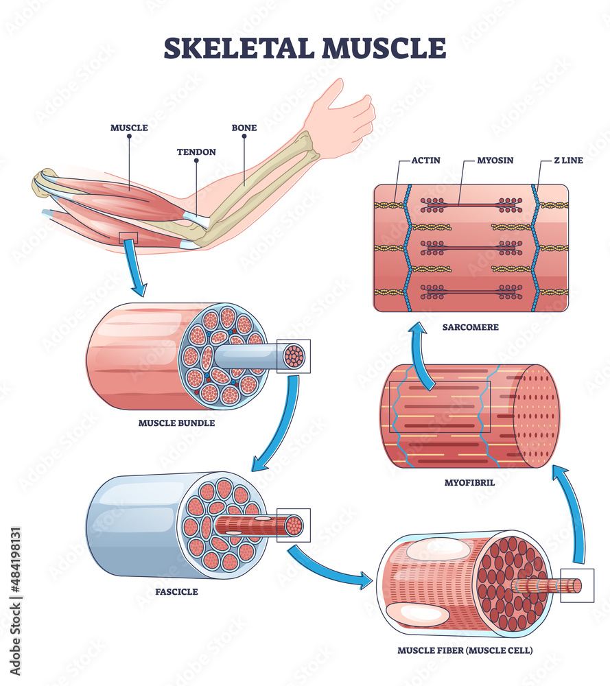
Skeletal muscle structure layers with anatomical arm closeups ...
Sarcomere (Muscle) Coloring - The Biology Corner The enter muscle fiber is surrounded by the sarcolemma (D), color this membrane brown. If expanded, the light and dark bands are shown as individual thick and thin filaments. Color the thick filaments (not labeled) red and the thin filaments blue. The Z line is the boundary between sarcomeres, named after its shape. Color the Z-line orange.

An optimized approach to study sub-sarcomere structure ...
Sarcomere Model Sarcomere Structure - YouTube This video was produced to help students of human anatomy at Modesto Junior College study our anatomical models.

4.1.3 Sarcomere Structure Diagram | Quizlet
Home Page: American Journal of Obstetrics & Gynecology Web04.08.2021 · AJOG's Editors have active research programs and, on occasion, publish work in the Journal. Editor/authors are masked to the peer review process and editorial decision-making of their own work and are not able to access this work in the online manuscript submission system.

Skeletal Muscle | Anatomy and Physiology I
(PDF) PERSONAL FITNESS TRAINER MANUAL - Academia.edu WebElliptical cross trainer (leg and arm exercise machine) has become popular for cardio respiratory fitness training. They are found in almost every gym house, recreation centres and hotels in Nigeria, all of which are imported as new products or fairly used.

Illustrations of (a) the sarcomere structure and (b) the ...
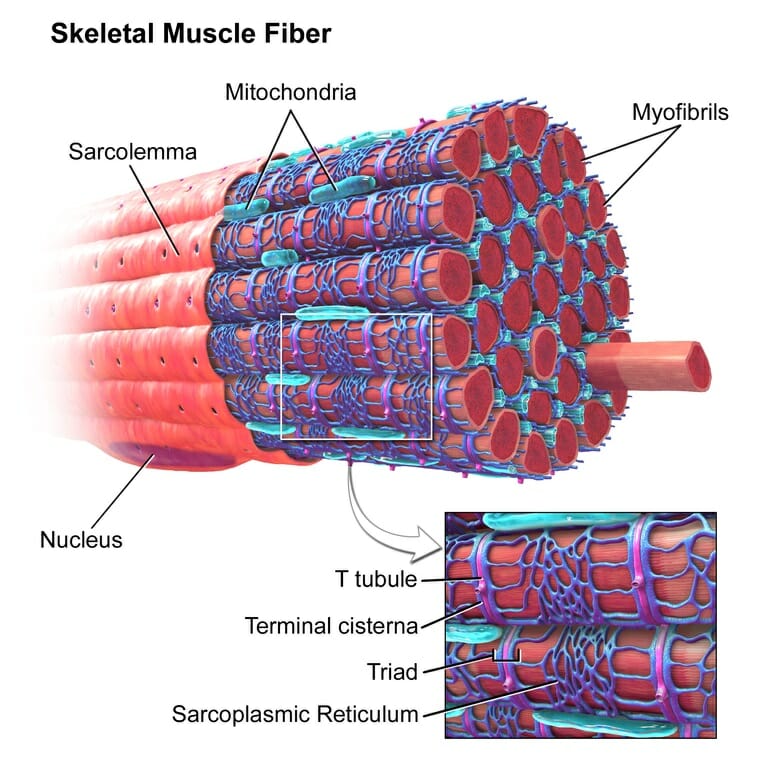
Skeletal Muscle Cell - an overview | ScienceDirect Topics WebSkeletal Muscle Cell. In cultured skeletal muscle cells and C2C12 myotubes, Ucn 2 inhibits insulin-induced Akt and ERK1/2 phosphorylation, consistent with the hypothesis that Ucn 2 functions as a local negative regulator of glucose uptake in skeletal muscle and suggests the possibility that suppression of the Ucn 2/CRH-R2 pathway may provide benefits in insulin …

Striated muscle sarcomere. a Schematic diagram showing the ...
Sarcomere | Definition, Structure, & Sliding Filament Theory - iBiologia Sarcomere Structure: Each sarcolemma or sarcomere is identical to biochemical composition to Plasmalemma, (that is another word for cell membrane). After observing under the microscope, a stacked pattern organized with a varied length of muscle fiber cells is seen. The bundle of filament arranged parallel to another is myofibril strands.

295 Sarcomere Images, Stock Photos & Vectors | Shutterstock
Titin - Wikipedia WebTitin / ˈ t aɪ t ɪ n / (contraction for Titan protein) (also called connectin) is a protein that in humans is encoded by the TTN gene. Titin is a giant protein, greater than 1 µm in length, that functions as a molecular spring that is responsible for the passive elasticity of muscle.It comprises 244 individually folded protein domains connected by unstructured peptide …

Myosin-binding protein C corrects an intrinsic inhomogeneity ...
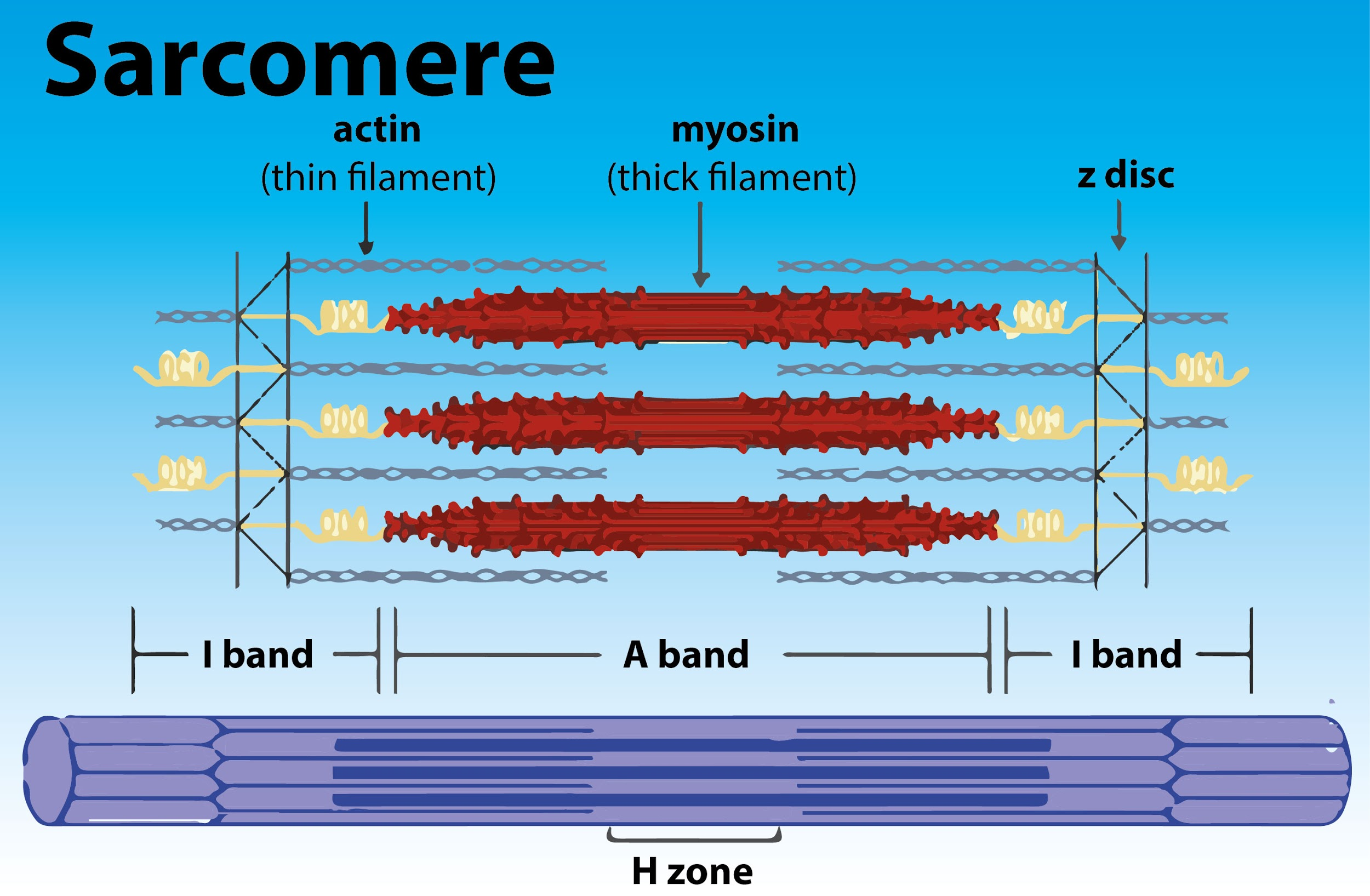
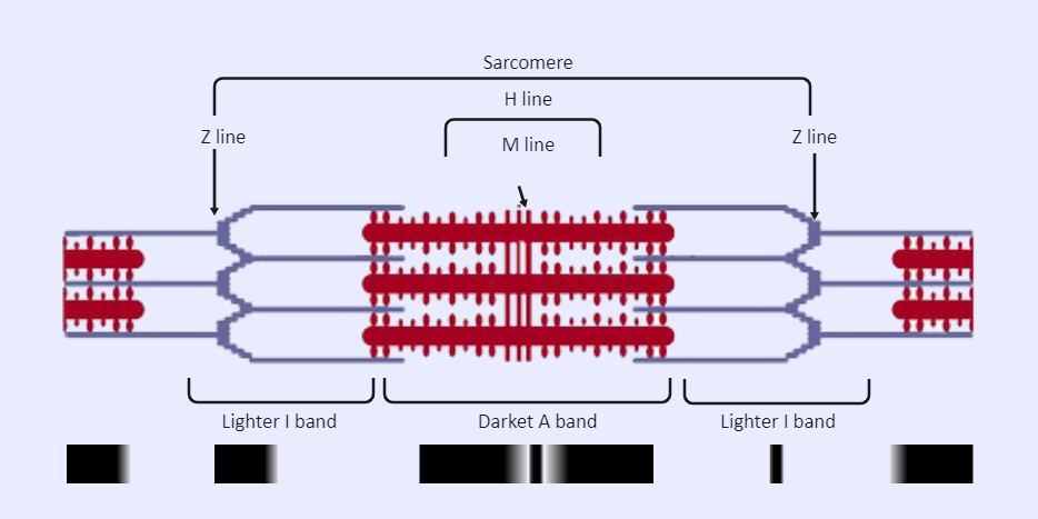
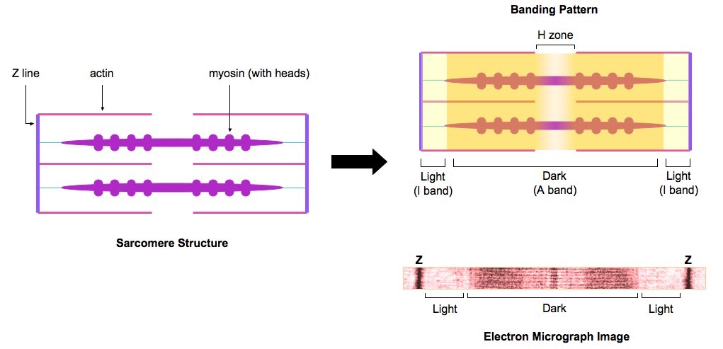
Sarcomere: anatomy, structure and function | Kenhub The structure of the sarcomere is traditionally described with dark and light bands visible under the microscope. This banding pattern in sarcomeres is due mainly to the arrangement of thick and thin myofilaments in each unit. These markings include: A bands (or anisotropic bands) - dark bands that contain whole thick filaments (myosin).

Draw and label a simple representation of two end to end ...
Home Page: Journal of Hand Surgery Web28.10.2022 · The Journal of Hand Surgery publishes original, peer-reviewed articles related to the pathophysiology, diagnosis, and treatment of diseases and conditions of the upper extremity; these include both clinical and basic science studies, along with case reports.Special features include Review Articles (including Current Concepts and The …

Human Physiology - Muscle
Sarcomere: Structure and Parts, Functions and Histology Although each sarcomere is small, several aggregated sarcomeres span the length of the muscle fiber. Myofilaments Each sarcomere consists of thick and thin bundles of the proteins mentioned above, which together are called myofilaments. By enlarging a portion of the myofilaments, the molecules that compose them can be identified.

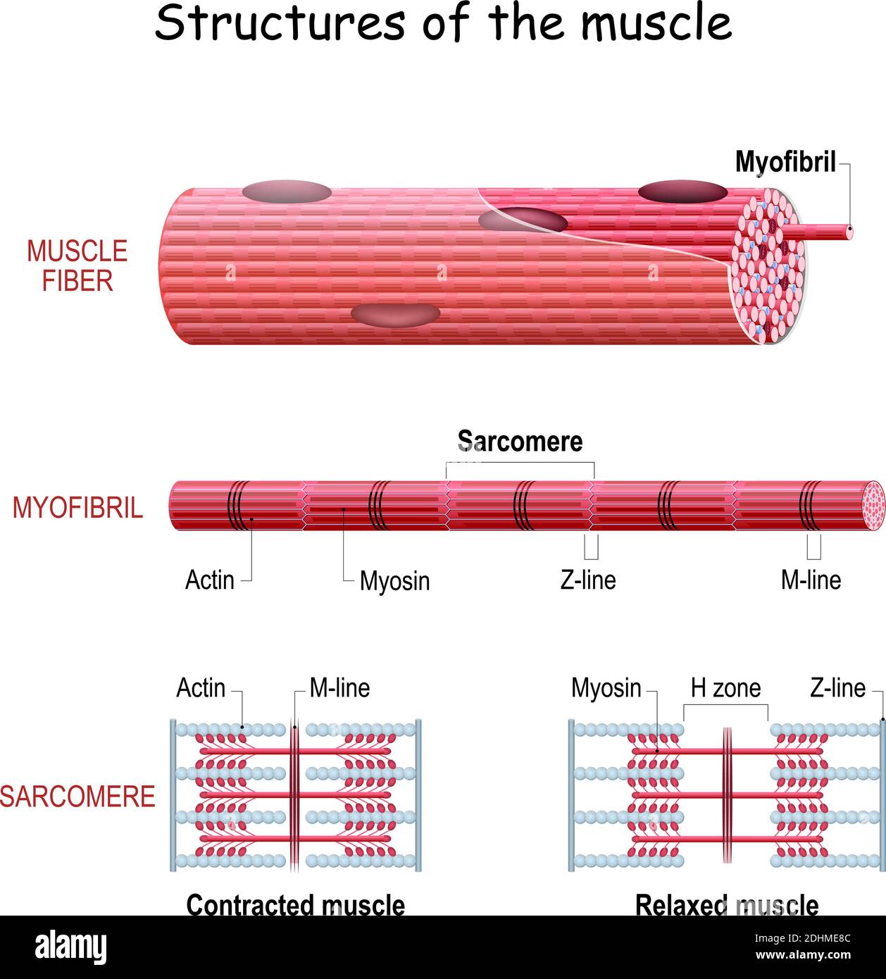
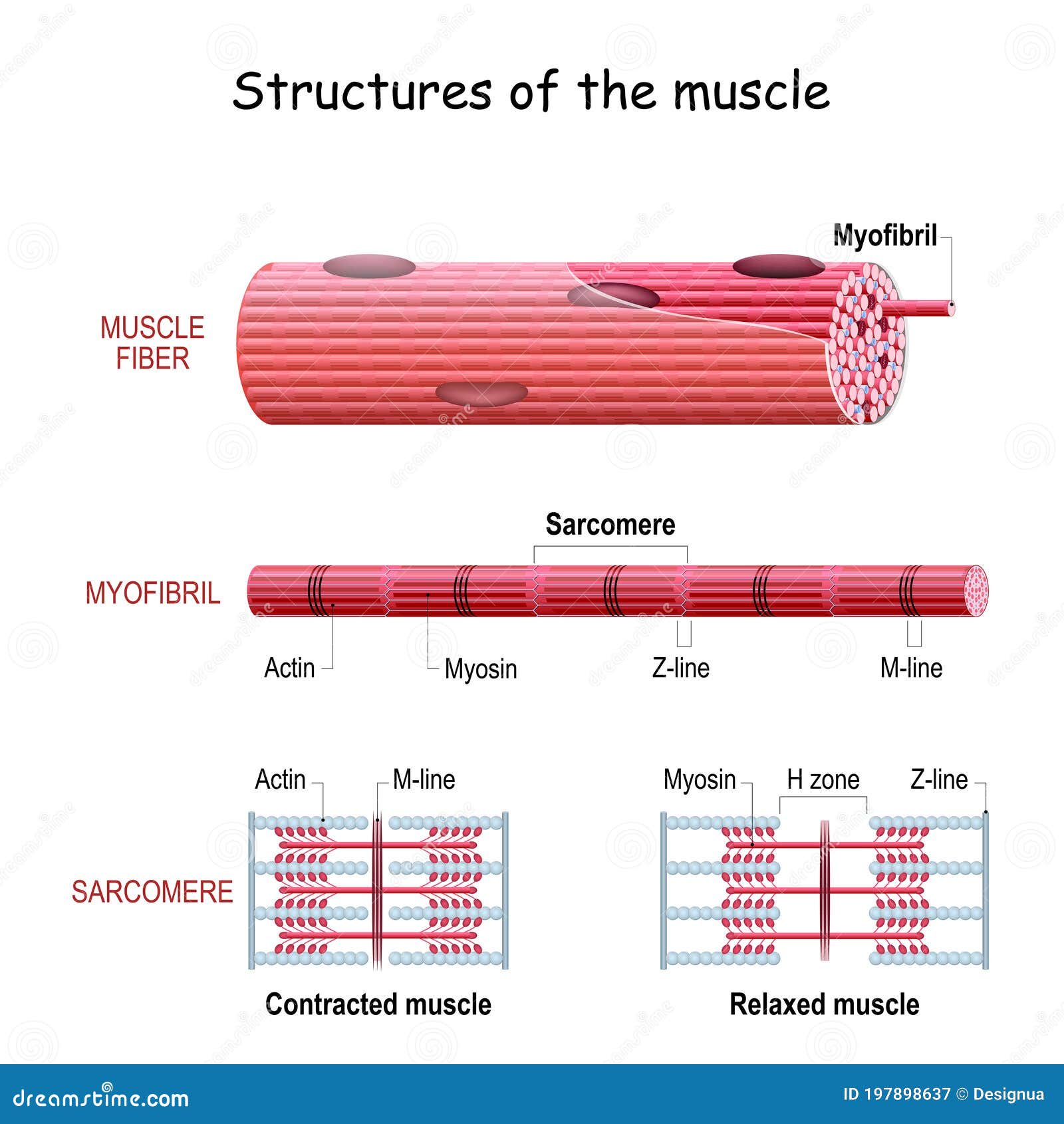
Structures of muscle with fiber, myofibril and sarcomere outline diagram
Sarcomere- Definition, Structure, Diagram, and Functions A sarcomere is a complex multicomponent biological system and functional unit of striated muscle which plays a vital role in transforming the chemical energy released upon the ATP hydrolysis into mechanical work. Skeletal muscles are made up of the basic unit called a sarcomere and all voluntary movement is initiated by this skeletal muscle.

Sarcomere structure | Anatomy and physiology, Physiology, Anatomy
en.wikipedia.org › wiki › EpimysiumEpimysium - Wikipedia Epimysium (plural epimysia) (Greek epi-for on, upon, or above + Greek mys for muscle) is the fibrous tissue envelope that surrounds skeletal muscle. It is a layer of dense irregular connective tissue which ensheaths the entire muscle and protects muscles from friction against other muscles and bones.

Muscle Structure And Control Of Contraction - Muscle System ...
Sarcomeres: "I" and "A" Bands, "M" and "Z" Lines, "H" Zone • A sarcomere is the basic contractile unit of skeletal muscle that is made of thick and thin filaments. • Thick filaments are organized bundles of myosin, while thin filaments are made of actin along with the two other regulatory proteins-troponin and tropomyosin. • Z-lines define the boundaries of each sarcomere.
Sarcomere - Definition, Structure, Function and Quiz ...
Glossary - The Cell - NCBI Bookshelf WebThe detection of radioisotopically labeled molecules by exposure to X-ray film. axonemal dynein. The type of dynein found in cilia and flagella. axoneme . The fundamental structure of cilia and flagella composed of a central pair of microtubules surrounded by nine microtubule doublets. β sheet. A sheetlike secondary structure of a polypeptide chain, …
Sarcomere - Wikipedia
Label the Sarcomere Structure Diagram | Quizlet Label the Sarcomere Structure Diagram | Quizlet Label the Sarcomere Structure + − Learn Test Match Created by jack_burton76 Plus Terms in this set (12) z disc ... mysosin (thick) ... thin (actin) filament ... I band ... A band ... I band ... H zone ... elastic (titin) filaments ... elastic (titin) filaments ... thin (actin) filament ...

Draw thediagram of a sarcomere of skeletal muscle showing ...
Sarcomere - Wikipedia A sarcomere (Greek σάρξ sarx "flesh", μέρος meros "part") is the smallest functional unit of striated muscle tissue. [1] It is the repeating unit between two Z-lines. Skeletal muscles are composed of tubular muscle cells (called muscle fibers or myofibers) which are formed during embryonic myogenesis. Muscle fibers contain numerous ...
Sarcomere Labeled | EdrawMax Template
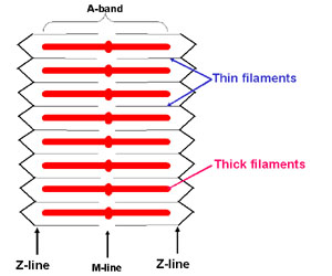
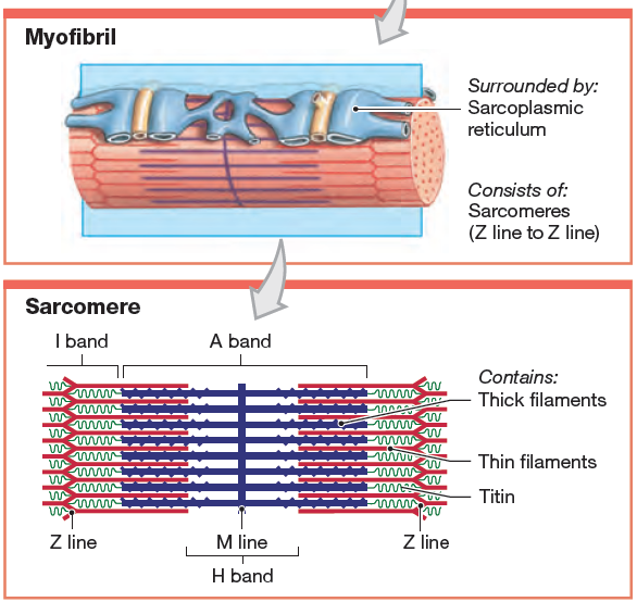
The Organization, Structure, and Function of Muscle The fundamental repeat unit within muscle that is responsible for contraction is the sarcomere. The sarcomere consists of a bundle of myosin-containing thick filaments flanked and interdigitated with bundles of actin-containing thin filaments (Fig. 1). The striated appearance of muscle results from the alternation of thick-filament-containing (A-Band) and thin-filament-containing (I-band ...
Sarcomere hi-res stock photography and images - Alamy
Diagram Of Sarcomere A sarcomere is the basic unit of striated muscle tissue. It is the repeating unit between two Z lines. Skeletal muscles are composed of tubular muscle cells which. sarcomere. Schematic: The Z line is depicted in black, myosin in red, actin in green/gray, and tropomyosin in blue. Image: MPI of Molecular Plant Physiology. Sarcomere definition.
Muscle: The Histology Guide
Metabolic characterization of hypertrophic cardiomyopathy in … Web09.05.2022 · Hypertrophic cardiomyopathy (HCM) is a common inherited cardiovascular disease with heterogeneous clinical presentations, governed by multiple molecular mechanisms. Metabolic perturbations ...

THE MUSCULOSKELETAL SYSTEM - Wellness for Body Mind Spirit
Page: American Journal of Obstetrics & Gynecology Aug 04, 2021 · AJOG's Editors have active research programs and, on occasion, publish work in the Journal. Editor/authors are masked to the peer review process and editorial decision-making of their own work and are not able to access this work in the online manuscript submission system.
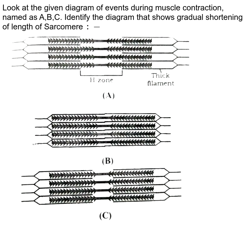
Look at the given diagram of events during muscle contraction ...
art-labeling activity: sarcomere structure - 4-h-dairy-posters Diagram and label a sarcomere including a thick filament thin filament A band H zone I band Z A. However thick and thin filamentsthe components of sarcomeresdo not shorten. Skin that has four layers of cells is referred to as thin skin From deep to superficial these layers are the. Solved Art Labeling Activity Sarcomere Structure Drag The Chegg ...

Identifying Regions in the Sarcomere
Epimysium - Wikipedia WebEpimysium (plural epimysia) (Greek epi-for on, upon, or above + Greek mys for muscle) is the fibrous tissue envelope that surrounds skeletal muscle. It is a layer of dense irregular connective tissue which ensheaths the entire muscle and protects muscles from friction against other muscles and bones. It is continuous with fascia and other connective tissue …
Sarcomeres | BioNinja
Sarcomere Structure: Actin & Myosin - Notes - NinjaNerd Medicine Sarcomere Structure: Actin & Myosin. Musculoskeletal. . Physiology. Get access to all our resources including notes and illustrations when you sign up to become a Ninja Nerd member. Become a Member. . View Notes.

Sarcomere Structure Diagram | Quizlet
Labeled Sarcomere Diagram A sarcomere is the basic unit of striated muscle tissue. It is the repeating unit between two Z lines. Skeletal muscles are composed of tubular muscle cells which. Sarcomeres are composed of thick filaments and thin filaments. The thin filaments Look at the diagram above and realize what happens as a muscle contracts.

9.1C: Sliding Filament Model of Contraction - Medicine LibreTexts
› books › NBK9926Glossary - The Cell - NCBI Bookshelf A sheetlike secondary structure of a polypeptide chain, formed by hydrogen bonding between amino acids located in different regions of the polypeptide. bacteriophage. A bacterial virus. basal body. A structure similar to a centriole that initiates the growth of axonemal microtubules and anchors cilia and flagella to the surface of the cell ...

Sarcomere Labeling Quiz
Skeletal Muscle | Anatomy and Physiology I

Draw the diagram of a sarcomere of skeletal muscle showing ...

Draw the diagram of a sarcomere of skeletal muscle showing ...

295 Sarcomere Images, Stock Photos & Vectors | Shutterstock

Sarcomere - an overview | ScienceDirect Topics
Second-harmonic microscopy of unstained living cardiac ...

Muscle structure – muscle under the microscope — Science ...

Art-labeling Activity: Sarcomere Structure Diagram | Quizlet

Structure Sarcomere Vector & Photo (Free Trial) | Bigstock
Structure Skeletal Muscle. Myofibril with Sarcomeres. Close ...
Sarcomere | Definition, Structure, & Sliding Filament Theory
Chapter 14 - Muscle Contraction
Post a Comment for "39 sarcomere structure labeled"